Step-by-step operative technique
A standard sequence done the same way every time is the single best protection against the day the anatomy is not standard.
Reviewed against its sources on 3 November 2026.
- Same sequence every time: position, four ports, fundus up and neck lateral, posterior peritoneum first, stay on the gallbladder wall.
- Clear the triangle from the gallbladder down until only two structures remain, then photograph the Critical View from both sides.
- No diathermy near the common duct, no clip on anything you cannot name, no blind clipping for bleeding.
01Principles before steps
- Identify, then divide. Nothing is clipped or cut until the Critical View of Safety (CVS) is obtained and confirmed in two views.
- Stay on the gallbladder. Dissection starts at the gallbladder wall and moves away from the bile duct, never toward it. The common duct does not need to be seen.
- Stop when the plan stops working. If the triangle cannot be cleared, change to a bail-out.
Sources and a note on the photographs
The operation described here follows the six strategies of the SAGES Safe Cholecystectomy Program (SAGES) and the safe steps in Tokyo Guidelines 2018 (Wakabayashi et al., 2018). Guideline The operative photographs on this page are reproduced from two open-access reviews by Gupta and colleagues under the Creative Commons BY-NC 4.0 licence, with the source and figure number under each; the labelled schematics are original to this site. Your own unit’s recorded operations are the best companion to both.
02Positioning, team and equipment
Patient
Supine, arms tucked or on boards. Reverse Trendelenburg 15 to 20 degrees and left tilt 10 to 15 degrees once ports are in.
Pregnancy after 20 weeks: left lateral tilt throughout (pregnancy)
Team
American: surgeon on the patient’s left, camera operator to the surgeon’s left, monitor over the right shoulder. French: surgeon between the legs.
Neither is safer; use what your unit teaches
Scope
30-degree, 10 mm. The angle is what shows the back of the triangle and the cystic plate.
A 0-degree scope makes the doublet view difficult
Instruments
Two atraumatic graspers, Maryland, hook or spatula, 5 mm clip applier (medium-large clips, 6 to 8 mm), scissors, suction-irrigator, retrieval bag.
Available: 10 mm clips, endoloop or suture for a wide duct, needle-holder for subtotal
Imaging
Cholangiography catheter and fluoroscopy, or the indocyanine green (ICG) camera, in the room if your unit uses them.
ICG is given at the right time for your regimen: hours before, or a low dose at induction or during the operation (dose table on the ICG page); how to perform and read a cholangiogram
Checklist
WHO surgical safety checklist, antibiotic prophylaxis where indicated (indications).
A named plan for the difficult case
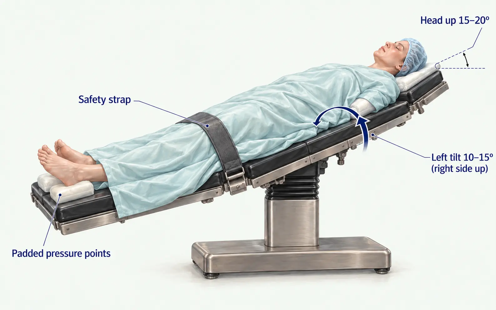
Image source and reuse
Before tilting, secure the patient, protect pressure areas and agree the change with the anaesthetist.
03Access and ports
- Open (Hasson), Veress or optical entry: no clear difference in major injury (Cochrane 2019); a trend to fewer failed entries with open. Use the method you were trained in. Contested
- Midline scar: avoid blind umbilical entry; use Palmer’s point.
- Insufflate to 12 to 15 mmHg; 10 to 12 is enough for most, and is used in pregnancy and poor cardiac function.
- Four ports are standard. The 2020 multi-society consensus prefers multi-port over single-incision because of higher bile duct injury and hernia rates in trials.
- Inspect the whole abdomen before touching the gallbladder.
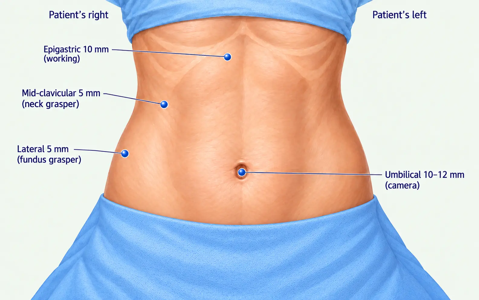
Image source and reuse
Sources
Entry techniques: Ahmad et al., Cochrane 2019. Multi-port over single-incision: Brunt et al., Ann Surg 2020. Guideline
Watch the operative demonstration
Watch Gupta’s official supplementary video on YouTube
Companion video to the 2023 technical article on achieving the critical view of safety, hosted by the Korean Association of Hepato-Biliary-Pancreatic Surgery. The article describes the author’s operative approach; it is a technical demonstration, not a separate guideline.
Opens on YouTube. Age-restricted surgical footage; YouTube may require sign-in. Read the accompanying article.
04The operation step by step
- Expose. Take down omental and duodenal adhesions with blunt traction, cold scissors or very brief diathermy on the gallbladder side. Neck adhesions are the inflammatory ones. Decompress a tense gallbladder with a needle through a port; send fluid for culture in cholecystitis.
- Retract. Fundus cephalad over the liver towards the right shoulder; Hartmann’s pouch laterally and downward towards the right iliac fossa. This opens the triangle and puts the cystic duct at a right angle to the common duct. Cephalad traction on the infundibulum lines the ducts up: the position of the classic injury.
- Place the landmarks. Rouvière’s sulcus, base of segment IV, duodenum, the duct if visible. Say them out loud. Stay above the sulcus-to-segment-IV line (anatomy).
- Open the peritoneum on the gallbladder, posterior leaf first. Start at the neck on the lateral (posterior) side, close to the wall, then the medial side. Opening the back first releases the neck, swings the pouch laterally and exposes the back of the triangle. Keep the incision high on the gallbladder, not on the duct.
- Clear the hepatocystic triangle. Sweep fat and lymphatics from the triangle towards the gallbladder, from the wall outward. The artery lies just deep to the lymph node; clear it for a short distance and no further. Diathermy sparingly, only on tissue lifted away from the ducts: 2 to 3 mm of spread from a hook can injure the common hepatic duct.
- Take the lower gallbladder off the cystic plate. Separate the lower third from the liver bed so the plate is seen behind the neck and the two structures from both sides. The step most often omitted, and the one that proves there is no third structure.
- Choose the order. Cystic plate first (separate the lower gallbladder early, alternating sides, then clear the triangle last; Gupta’s preference for leverage and distance from the hilum) or Calot’s triangle first (duct and artery first, plate afterwards). Hybrids are common. The Critical View is not complete until the plate is seen. Expert opinion
- Confirm the CVS and pause. Three criteria, anterior and posterior views (the doublet view), images recorded. Say “CVS achieved, two structures, clipping the cystic duct” and let anyone challenge it. Cholangiography or ICG now, before clipping (CVS page).
- Clip and divide the cystic artery. Two clips on the stay side, one on the gallbladder side; divide with scissors, not diathermy. Artery before duct where it lies anterior to the duct.
- Clip and divide the cystic duct. Clips near the gallbladder, a stump of a few millimetres; do not chase the duct to the junction. Two plus one; jaws fully closed, nothing else caught. Wide (over 6 to 8 mm) or thick duct: loop, suture or stapler, not stacked clips, and ask why it is wide (duct stones, Mirizzi).
- Dissect the gallbladder off the liver. Neck up and lateral; hook or spatula in the plane between wall and cystic plate, areolar tissue in view, alternating traction. Going deep bleeds and divides subvesical ducts. Inspect the bed and clips before the last attachment is divided.
- Check, wash, extract. Drop the pressure to unmask venous bleeding. Irrigate after spillage and retrieve stones. Bag the gallbladder; enlarge the fascia rather than tear the bag. Record spilled stones and tell the patient (complications).
- Drain? Not routinely: more wound infection, no fewer collections (Cochrane 2013). Reasonable after subtotal cholecystectomy, an inflamed bed or a suspected small leak; never a substitute for dealing with a known leak.
- Close. Fascia at every port of 10 mm or more, especially the umbilicus, under vision with absorbable suture. Local anaesthetic to the port sites (day case). Absorbable sutures or glue to skin.
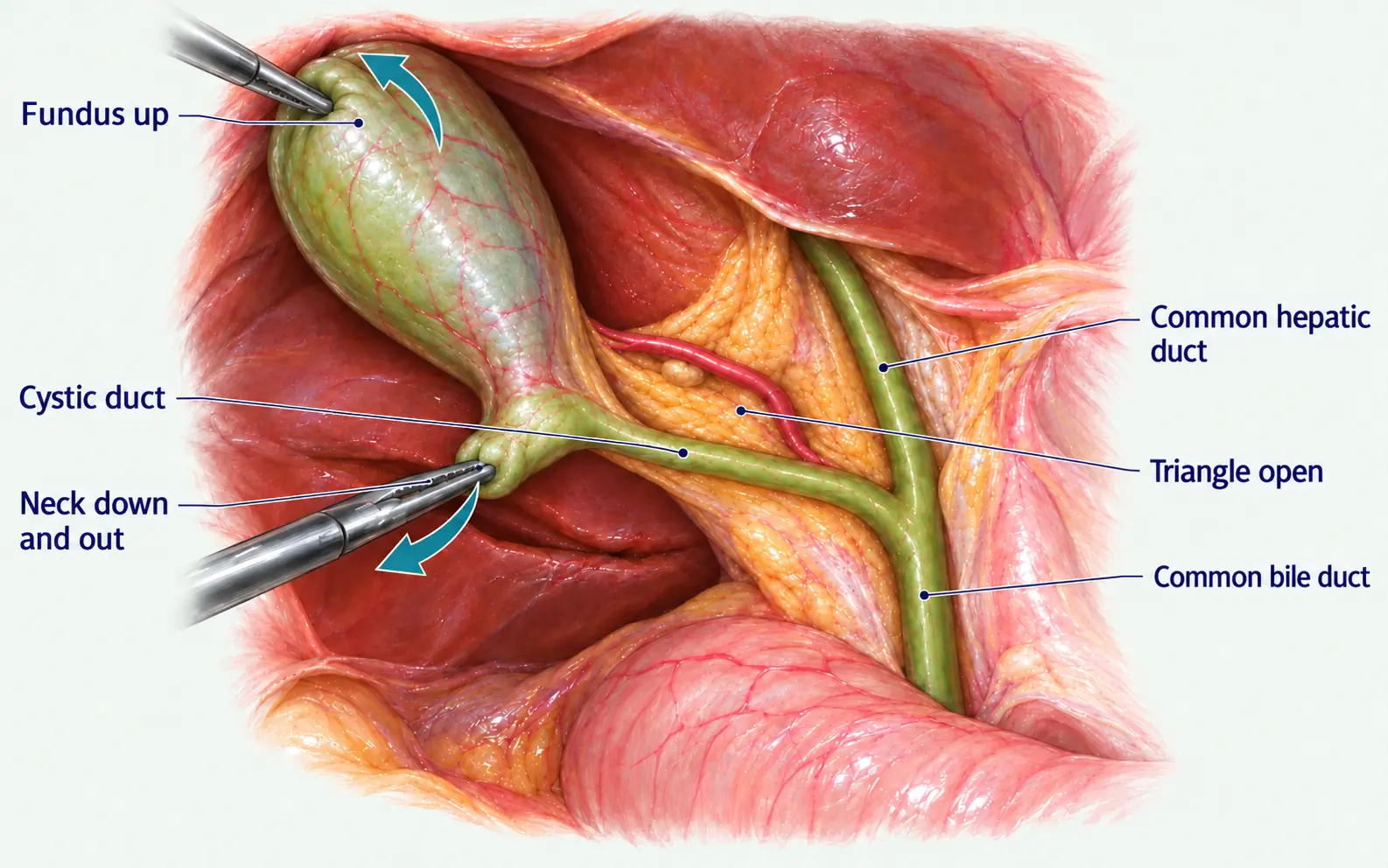
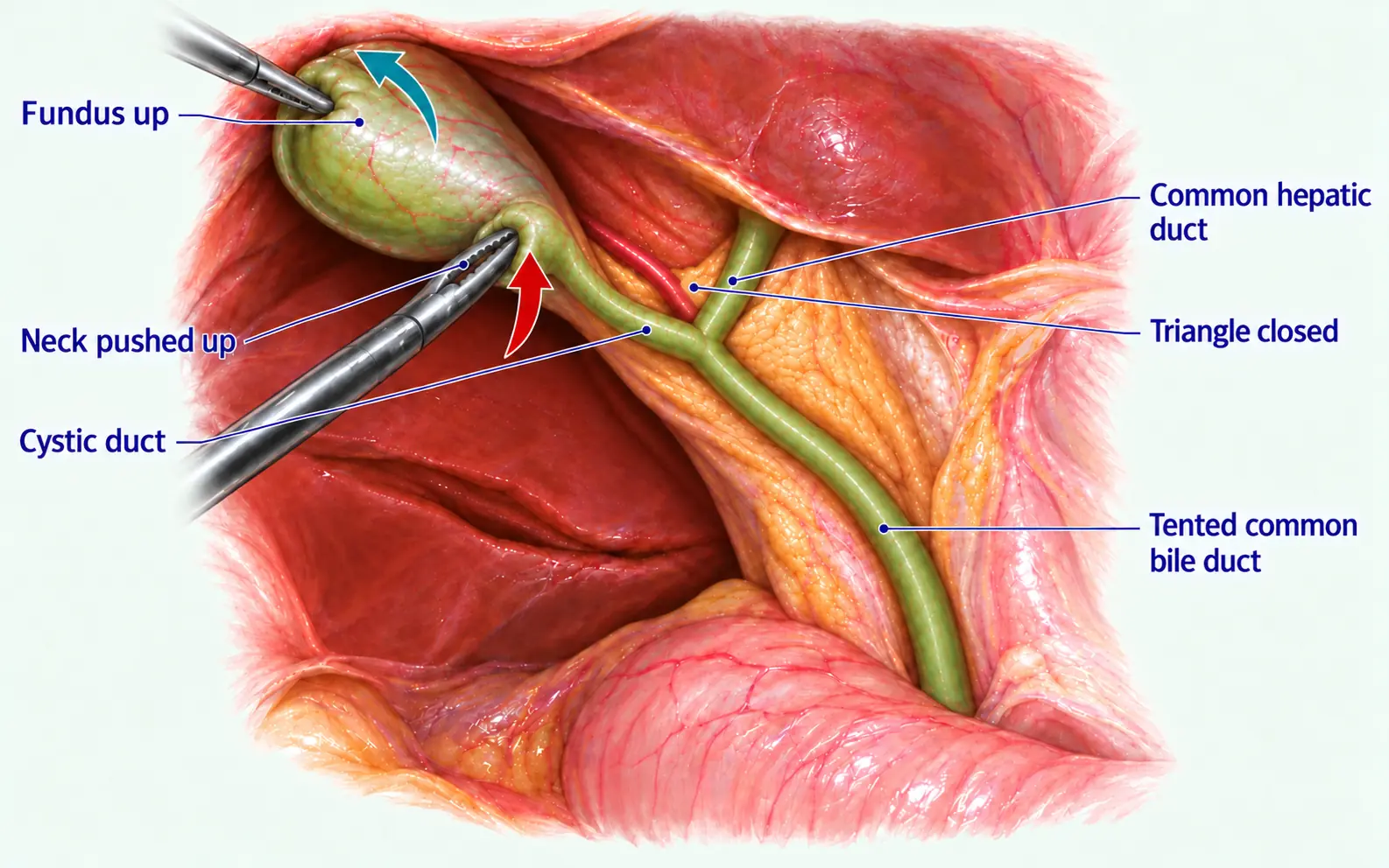
Image source and reuse
Two views of correct two-handed retraction and one of fundal retraction alone. Click any photograph to see it full screen.
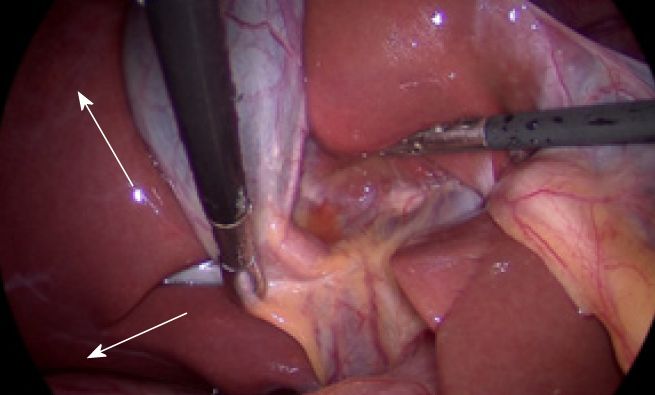
Image source and reuse
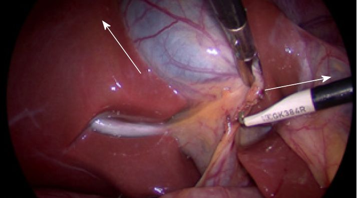
Image source and reuse
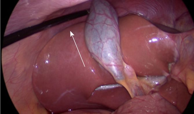
Image source and reuse
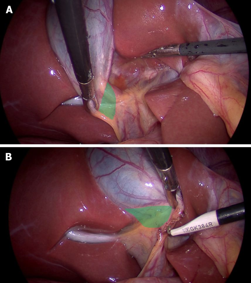
Image source and reuse
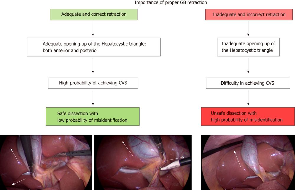
Image source and reuse
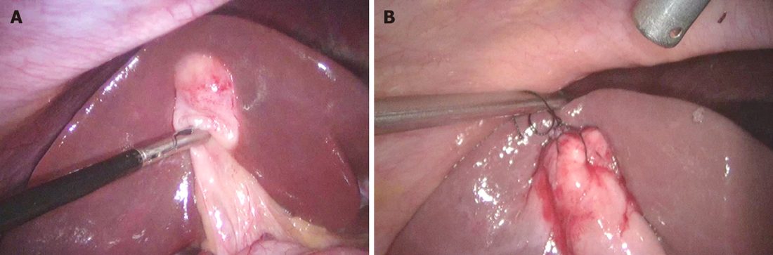
Image source and reuse
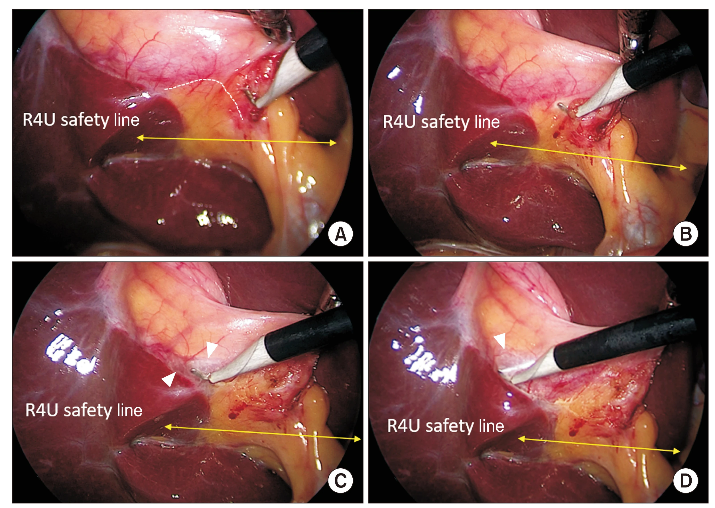
Image source and reuse
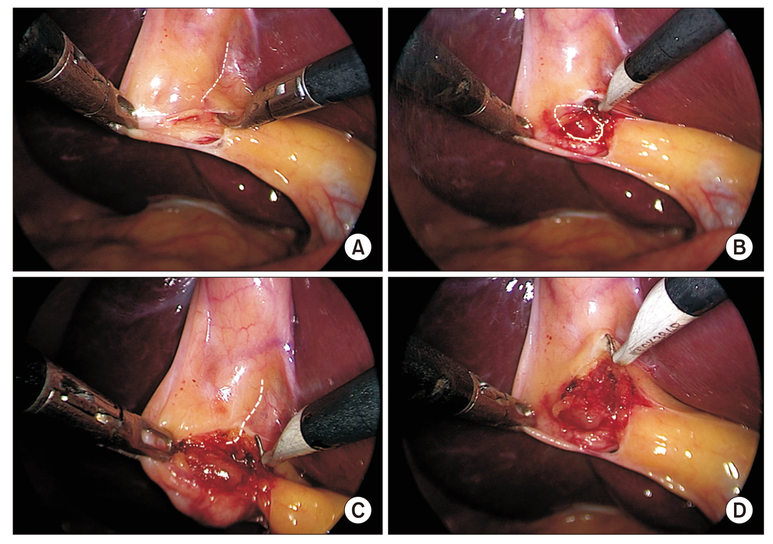
Image source and reuse
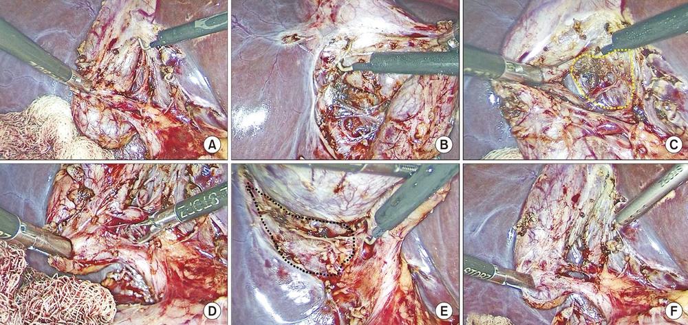
Image source and reuse
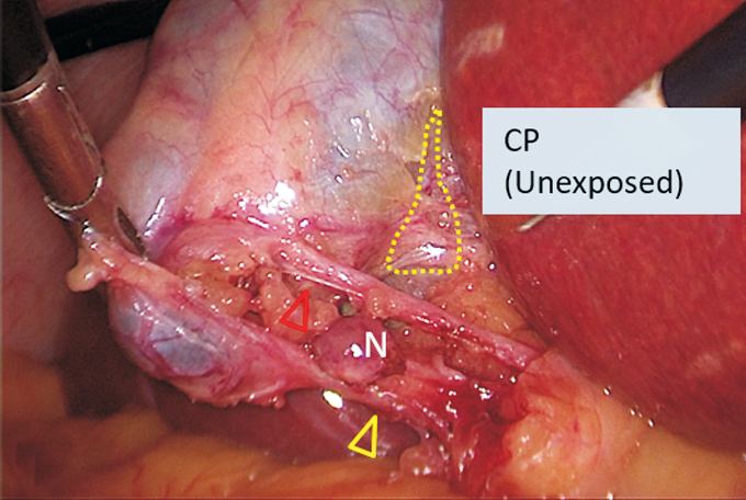
Image source and reuse
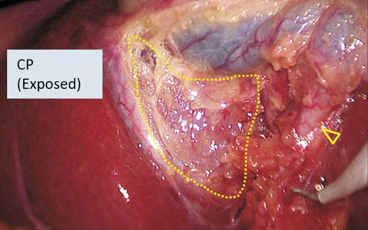
Image source and reuse
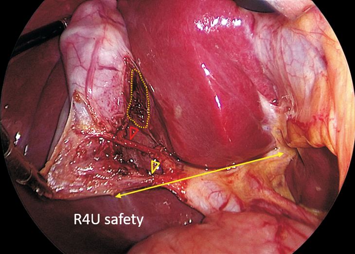
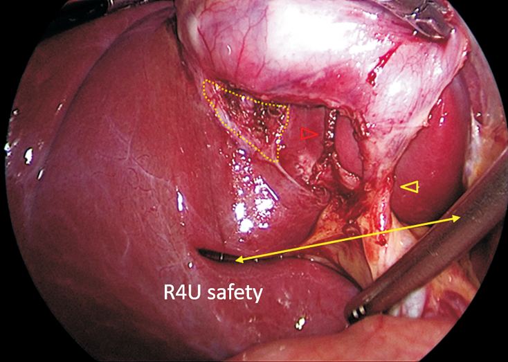
Image source and reuse
Reproduced from Gupta V. How to achieve the critical view of safety for safe laparoscopic cholecystectomy: Technical aspects. Ann Hepatobiliary Pancreat Surg 2023;27(2):201-210, Fig. 8B (doi:10.14701/ahbps.22-064), under the CC BY-NC 4.0 licence. No changes other than resizing.
Before clipping: confirm all three CVS criteria in the real anterior and posterior views above, then pause with the team. A photograph of applied clips alone does not document the preceding CVS.
More detail and sources
Retraction. Cephalad traction on the infundibulum lines up the cystic and common ducts and is the position in which the classic injury happens; lateral and downward traction on Hartmann’s pouch opens the hepatocystic triangle (Gupta and Jain 2019).
Dissection order. Gupta describes two sequences. In the cystic plate first approach the lower gallbladder is separated from the liver bed early, alternating between the lateral and medial sides until at least the lower third of the plate is exposed, and the triangle is cleared last; the authors prefer it because it gives more leverage on the gallbladder and keeps the dissection away from the hilum. In the Calot’s triangle first approach the duct and artery are delineated first and the plate exposed afterwards (Gupta, Ann Hepatobiliary Pancreat Surg 2023). Expert opinion The preference for the plate-first order is the authors’ practice, not a trial finding.
Doublet view. Sanford and Strasberg (J Am Coll Surg 2014). The intra-operative momentary pause is part of the SAGES programme.
Drains. Routine drainage is not supported after uncomplicated laparoscopic cholecystectomy; reserve a drain for a specific clinical indication (Gurusamy et al., Cochrane 2013). Systematic review
05Energy and clips
- Short bursts of monopolar coagulation on tissue tented away from what lies behind it.
- Consider bipolar or ultrasonic devices in a fibrotic bed: less lateral spread, same identification steps.
- Apply the six clip rule: two structures, three clips each. Reaching for a seventh? Ask what you are clipping. Expert opinion
- Use diathermy inside the triangle before the structures are identified.
- Use it on or next to the bile duct.
- Stack clips on a thick or wide duct, or let a clip rest against the common duct: leaks now, strictures later.
Sources
Six clip rule: Deng et al., Art Surg 2021. Expert opinion
06The operation note
Write the note as evidence. “CVS obtained” without a description is worth little when the patient returns jaundiced.
- Indication and context; findings and difficulty grade.
- Landmarks seen; how the CVS was obtained and documented; variants.
- Cholangiography or fluorescence findings.
- Number and position of clips; spillage; drains; plan.
“Posterior and anterior peritoneum opened at the neck; hepatocystic triangle cleared of fat; lower third of gallbladder separated from the cystic plate; two structures only seen entering the gallbladder, confirmed in anterior and posterior views (images saved). Pause, confirmed by team. Cystic artery (2 + 1 clips) then cystic duct (2 + 1 clips) divided. No spillage. Fascia closed at umbilicus.”
Source
WSES recommends that the note records the indication and clinical context, the operative findings and difficulty grade, the landmarks seen, whether and how the Critical View of Safety was obtained and documented, any anatomical variants, the findings of cholangiography or fluorescence, the number of clips and their position, any spillage, drains and the plan (de’Angelis et al., WSES 2021). Guideline
07Closing the cystic duct and retrieving the gallbladder
Clips, ligatures or loops
Metal clips are standard. Absorbable clips and absorbable ligatures were no safer and no more dangerous in three small trials; ligating took about 12 minutes longer. A wide or thick duct gets a loop, a suture or a stapler, as above.
Cochrane 2010: 3 trials, 255 patients, evidence too thin to prefer one method
Clip migration
A clip in the common duct years later, acting as a stone nidus, is reported in case series only. It is rare. Keep clips near the gallbladder, and keep the number down. Expert opinion
Retrieval bag
Use a bag when the gallbladder is perforated, infected, thick-walled or suspicious, or when stones have spilled. Many surgeons use one every time. It means a slightly larger fascial opening.
Meta-analysis, 8 studies, 1,805 patients: fewer wound infections and less bile spillage with a bag; no difference in port-site hernia
Spilled stones: retrieve what you can see, wash, record the spillage in the note, and tell the patient. Lost stones cause abscesses months later. See spilled gallstones.
Sources
Gurusamy et al., Cochrane 2010: absorbable versus non-absorbable clips (2 trials, 150) and absorbable ligatures versus clips (1 trial, 105); morbidity no different; all trials at high risk of bias. Systematic review Mohamedahmed, Cureus 2023: four randomised and four non-randomised studies; intra-abdominal collections similar. Mixed trial and observational data
Test yourself further: Practise questions on Operative technique in the Exam zone.