Predicting and grading difficulty
Difficulty can be predicted, graded and planned for. Grading also gives you a shared language for the operation note, the handover and the audit.
Reviewed against its sources on 25 October 2026.
- Predict difficulty before the operation and plan senior help, imaging and a bail-out.
- Grade every case with one scale (Parkland, Nassar or G10) and record it.
- Warning signs outrank any score: a triangle that will not clear, a structure you cannot name, no progress after 30 minutes.
01Why grade difficulty
- Plan: the right surgeon, the right list, the right conversation with the patient.
- Compare: a unit’s conversion or subtotal rate means nothing without the case mix.
- Communicate: a vocabulary in the note that a hepatobiliary surgeon reading it later can trust.
Source
The 2020 multi-society consensus suggests grading acute cholecystitis with Tokyo 2018, AAST or another validated scale, and considering the listed difficulty factors when planning (Brunt et al., Ann Surg 2020). Guideline
02Pre-operative predictors
| Where | Predictors of a difficult operation |
|---|---|
| CholeS (Nassar 2020) | Older age; higher ASA; male sex; duct stones or cholecystitis rather than colic; thick-walled gallbladder; dilated duct; previous endoscopic retrograde cholangiopancreatography (ERCP); non-elective operation |
| History | Previous admission with cholecystitis (especially more than one); symptoms over 72 hours; previous cholecystostomy; previous upper abdominal surgery; BMI over 30; diabetes; cirrhosis |
| Examination | A palpable mass; a very tender right upper quadrant with guarding |
| Bloods | White cells above 18,000/mm³; CRP above 10 to 15 mg/dL |
| Ultrasound | Wall over 4 to 5 mm; pericholecystic fluid; contracted gallbladder with stones; impacted stone in the neck; stone over 2 cm; dilated duct; intrahepatic position |
Indian pre-operative scores (such as Randhawa and Pujahari) need only bedside and ultrasound findings and are widely taught for DNB and MS, but are validated only in single-centre series. Contested The UK CLOC score predicts conversion specifically.
More detail and sources
Nassar and colleagues derived and validated a score in over 8,000 patients from the CholeS data, with difficulty defined as Nassar grade 3 to 5 at operation; 11 percent of low-risk and 80 percent of high-risk patients had a difficult operation, with an area under the curve of 0.79 on external validation (Nassar et al., Surg Endosc 2020). Observational The Randhawa and Pujahari score combines age, sex, history of hospitalisation, body mass index, abdominal scar, palpable gallbladder, wall thickness, pericholecystic collection and impacted stone. The CLOC score (age, sex, indication, ASA, duct diameter and wall thickness) is from Sutcliffe et al., HPB 2016.
03Intra-operative grading scales
Three scales measure different things. Parkland grades inflammation at first view. Nassar grades the whole field: gallbladder, pedicle and adhesions. G10 scores ten findings that predict conversion. For a general unit: Parkland at first view plus Nassar at the end, recorded in the note.
Parkland (scored at first view, before dissection)
| Grade | Findings |
|---|---|
| 1 | Normal gallbladder, no adhesions |
| 2 | Minor adhesions at the neck, otherwise normal |
| 3 | Any of: hyperaemia, pericholecystic fluid, adhesions to the body, distended gallbladder |
| 4 | Any of: adhesions obscuring most of the gallbladder; grade 1 to 3 with abnormal liver anatomy, intrahepatic gallbladder or impacted stone (Mirizzi) |
| 5 | Any of: perforation, necrosis, gallbladder cannot be seen for adhesions |
Predicts longer operations, conversion, subtotal, leak and complications. Weakness: it says little about the hepatocystic triangle, where the danger lies.
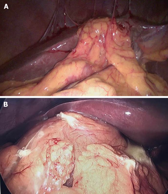
Image source and reuse
Nassar (worst of three components)
| Grade | Gallbladder | Cystic pedicle | Adhesions |
|---|---|---|---|
| 1 | Floppy, non-adherent | Thin and clear | Simple, up to the neck |
| 2 | Mucocele, packed with stones | Fat-laden | Simple, up to the body |
| 3 | Deep fossa, acute cholecystitis, contracted, fibrosis, pouch adherent to the common bile duct (CBD), impacted stone | Short, dense or obscured | Dense, up to the fundus; involving flexure or duodenum |
| 4 | Completely obscured | Impossible to clarify | Dense, fibrous, wrapping the gallbladder; duodenum or flexure hard to separate |
| 5 | Mirizzi type 2 or above, cholecysto-enteric or cholecysto-biliary fistula, or complete obliteration of the triangle | ||
Grades 3 to 5 are “difficult”. In AMBROSE (21,706 patients) the Nassar grade was one of the five strongest predictors of 30-day complications. It is the scale that best describes the triangle, so this site uses it in the note template.
G10 (WSES, revised 2019)
- Changed recently Name the version you used (2015 or 2019). The 2015 and 2019 scores share a name and range but not the same items: the 90-minute item was dropped in 2019 and replaced by access limited by adhesions.
- The adhesion categories are alternatives: choose one, do not add them.
- A low score early protects nobody against a hostile triangle found later. The score informs the plan; it never overrides the stop rule.
Tokyo Guidelines 2018 difficulty findings
A Japan-Korea-Taiwan Delphi panel agreed 25 findings, grouped by location: around the gallbladder, in the hepatocystic triangle, in the gallbladder bed, and outside the gallbladder. The two most likely to lead to injury: fibrosis in the triangle and fusion of the neck to the common duct.
More detail and sources
A 2025 study applied all three scales to elective and emergency cases and found higher grades on all three in emergency operations, with the scales broadly agreeing (Narvaez Gonzalez, Cureus 2025). Parkland: Madni et al., Am J Surg 2018, validated in several countries. Nassar: first published in 1995 and extended with grade 5 for the CholeS analysis (Griffiths et al., Surg Endosc 2019); predictor of complications in AMBROSE Collaborative, Ann Surg 2025.
G10. The original 2015 score graded the gallbladder (adhesions covering less than half; more than half; distended or contracted; unable to grasp with atraumatic forceps; a stone of 1 cm or more impacted in Hartmann’s pouch) and five additional factors (BMI over 30; adhesions from previous surgery limiting access; sepsis or cholecystitis; bile or pus outside the gallbladder; time to identify the cystic artery and duct over 90 minutes), with 0 to 1 mild, 2 to 4 moderate, 5 to 7 severe, 8 to 10 extreme (Sugrue et al., World J Emerg Surg 2015). The revised 2019 score was validated prospectively by WSES in 504 patients from 16 countries; 71 (14 percent) were converted, and a score of 3 or more best predicted conversion, with an area under the curve of 0.77 (Sugrue et al., World J Emerg Surg 2019). A 2024 validation from Nepal found similar discrimination with a locally different cut-off (Baral et al., 2024). Contested
TG18 findings. Around the gallbladder: adhesions, scarring, a fibrotic or necrotic wall, an impacted stone, a contracted gallbladder, an atrophic gallbladder buried in the liver. In the triangle: fibrosis, fat, scarring, inability to identify the cystic duct, fusion of the neck to the common duct. In the bed: fibrosis, bleeding, an intrahepatic gallbladder. Outside: adhesions to duodenum or colon, obesity, cirrhosis, previous surgery (Iwashita et al., J Hepatobiliary Pancreat Sci 2017). Guideline
04Intra-operative warning signs
Scores describe the field. These signs describe the moment the operation turns dangerous. Each one triggers the stop rule on the bail-out page.
No landmarks
Rouvière’s sulcus and the base of segment IV cannot be found, or the duodenum is stuck to the neck.
The neck does not move
Lateral traction on Hartmann’s pouch does not open the triangle: the neck is fused to the ligament.
The shield
Stiff peritoneum over the triangle that will not peel (the shield of McElmoyle): chronic fibrosis.
A duct bigger than expected
A “cystic duct” over 5 to 6 mm, one that keeps going without a junction, or one a medium-large clip cannot close, is the common duct until proven otherwise.
Vanishing Calot syndrome: the only tube in view runs vertically below the R4U line and behind the duodenum. It is the common bile duct
A third structure
A second duct or a third structure after the first two are clipped.
Bile where it should not be
Bile from the hepatoduodenal ligament during dissection means a duct has been opened.
Bleeding in the triangle
Pulsatile bleeding suggests the right hepatic artery. Blind clipping or diathermy here is the mechanism of vasculobiliary injury.
Progress has stopped
Thirty minutes with no change in the view, or dissection going deeper rather than clearer. Expert opinion
Your own state
Irritation, speed, and the sentence “I just need to get round this” (culture of safety).
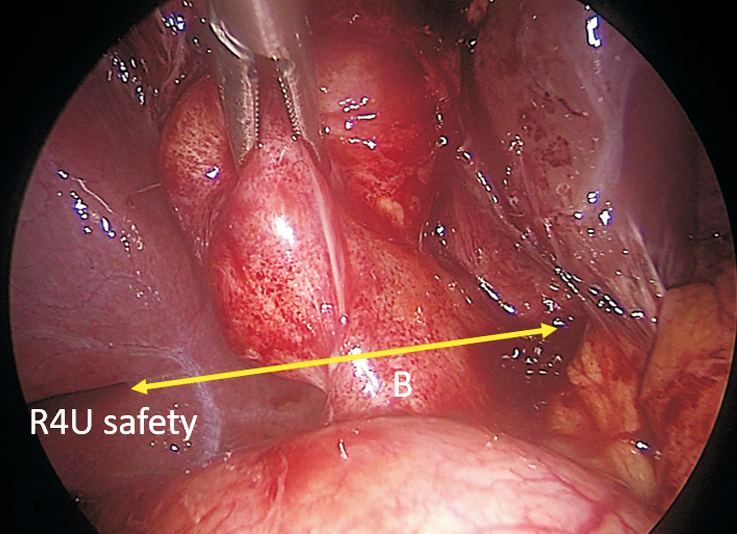
Image source and reuse
Difficulty is predictable from the ultrasound and the history, and recognisable in the first five minutes. Decide before the first clip whether this operation can be finished safely with a Critical View of Safety. If the answer is “probably not”, change the plan now, not after an hour of bleeding.
Sources
Duct calibre rule: Gupta and Jain, World J Gastrointest Surg 2019. Vanishing Calot syndrome: Gupta, Ann Hepatobiliary Pancreat Surg 2023. The Tokyo surgical management paper suggests that if the CVS has not been obtained within a reasonable time the surgeon should move to a bail-out rather than persist. Expert opinion
Test yourself further: Practise questions on Predicting difficulty in the Exam zone.