Special situations
The standard operation needs changing in a handful of situations. Knowing which ones, and how, is what separates a safe surgeon from a lucky one.
Reviewed against its sources on 3 November 2026.
- Cirrhosis: operate for symptoms in Child A and selected B, expect bleeding, avoid Child C; a subtotal is often the safe operation.
- Pregnancy: treat acute cholecystitis promptly with obstetric input. For third-trimester biliary colic, individualise surgery versus observation.
- Mirizzi needs fundus-first or subtotal with the duct protected; suspected cancer needs staging and a hepatobiliary unit.
01Cirrhosis and portal hypertension
- Gallstones are twice as common in cirrhosis. More bleeding, conversion, decompensation and death; risk tracks Child-Pugh class and MELD, not cirrhosis as such.
- Laparoscopy is the approach of choice in Child A and selected B (WSES 2020 for acute cholecystitis). Trials: fewer complications, less infection, shorter stay than open.
- Child C: no elective cholecystectomy. Cholecystitis: antibiotics, percutaneous or endoscopic drainage, and a conversation with the transplant unit.
- Open umbilical entry away from a recanalised umbilical vein; blood available; correct coagulopathy.
- Expect liver bed bleeding, varices in the ligament and a liver that will not retract.
- Consider leaving the posterior wall on the liver during subtotal cholecystectomy rather than dissecting a bleeding fossa. An open versus closed remnant defines fenestrating versus reconstituting subtotal.
- Drains selectively: ascites leaks through drain sites. Watch for decompensation afterwards.
- Go fundus-first into a hard liver.
- Operate electively on a Child C patient.
Sources
Pisano et al., WJES 2020 Guideline; meta-analysis of randomised trials, laparoscopic versus open in cirrhosis (de Goede et al., Br J Surg 2013).
02Pregnancy
- For acute cholecystitis, plan early laparoscopic cholecystectomy with obstetric and anaesthetic input; pregnancy alone is not a reason to defer necessary treatment.
- For recurrent biliary symptoms, discuss surgery during pregnancy, balancing recurrence, disease severity and gestational age.
- For third-trimester biliary colic, either surgery or non-operative management may be appropriate; agree a plan with the patient and obstetric team.
- Technique: left lateral tilt after 20 weeks; open entry or an entry point adjusted to fundal height; 10 to 15 mmHg; monitor end-tidal CO2; fetal heart before and after; mechanical thromboprophylaxis; obstetric input.
- Cholangiography is safe with uterine shielding and brief screening; SAGES 2025 lists pregnancy as a reason not to do a routine one. Indocyanine green (ICG) is not licensed in pregnancy. Endoscopic retrograde cholangiopancreatography (ERCP) when needed, with shielding.
Sources
Kumar et al., SAGES pregnancy guideline 2024: conditional recommendations favour laparoscopic cholecystectomy for biliary disease (very low certainty) and specifically acute cholecystitis (low certainty). For third-trimester biliary colic, either operative or non-operative management may be considered (expert opinion). The trimester comparison below is observational and cannot establish that delaying an individual patient’s operation improves outcomes. Technical considerations also draw on Pearl et al., SAGES 2017. Guideline; trimester outcomes (Cheng et al., J Am Coll Surg 2021) Observational, debated; Kumar et al., Surg Endosc 2025.
03Mirizzi syndrome
Obstruction of the common hepatic duct by a stone impacted in the neck or cystic duct: by compression and inflammation (Csendes type I) or because the stone has eroded into the duct (types II to IV, a cholecystobiliary fistula); type V adds a cholecystoenteric fistula.
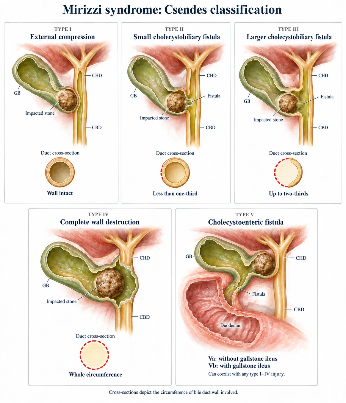
Illustration sources
Suspect it
Jaundice with a thick-walled contracted gallbladder and a large stone at the neck; common hepatic duct dilated above the gallbladder and normal below; on magnetic resonance cholangiopancreatography (MRCP) a stone at the junction.
On the table
Neck fused to the ligament; “cystic duct” very short or absent; no triangle to display.
Stop-rule situation: do not dig the stone out of the duct, do not clip what looks like a cystic duct
Type I
Cholecystectomy, often subtotal, with cholangiography to confirm an intact duct. Laparoscopy is reasonable with experience.
Types II to IV
Subtotal cholecystectomy leaving a cuff to close the fistula over a T-tube, or hepaticojejunostomy for larger defects. These are hepatobiliary operations, best done by or with a hepatobiliary team. Pre-operative ERCP and stenting diagnoses and protects the duct, and is sometimes definitive. Expert opinion and case series
Source
04Gallbladder perforation and gangrene
- Gangrene in up to 20 percent of acute cholecystectomies in the elderly and diabetic; free perforation is rarer.
- Niemeier: I free perforation with biliary peritonitis; II localised with pericholecystic abscess; III perforation into a viscus (cholecystoenteric fistula).
- Warning: sicker than the findings suggest; white cells above 15,000 to 18,000/mm³; rising CRP; on CT, wall discontinuity, gas, absent enhancement. These are Tokyo II or III: surgery or drainage urgently. Antibiotics alone will not control a gangrenous gallbladder.
- At operation: decompress with a needle before grasping; the wall tears, stones spill, the duct is friable; expect a subtotal; cultures; wash out.
- Type III is usually chronic. Gallstone ileus: enterolithotomy; leave the fistula and gallbladder at the first operation.
05Incidental gallbladder cancer
Found in about 0.2 to 2 percent of specimens; commoner in north India, Chile and eastern Europe. There are three situations, depending on when the cancer is found.
1. Suspected before
A mass, focal thickening, a polyp of 10 mm or more or growing, or a porcelain gallbladder with other findings: cross-sectional imaging and a hepatobiliary discussion first.
No laparoscopic cholecystectomy on a suspected cancer: spillage seeds peritoneum and ports
2. Suspected during
Hard, irregular, fixed gallbladder; thick white wall; nodes; liver invasion. Stop. Do not perforate, do not do a partial through tumour, biopsy only if it changes the next step and will not spill, photograph, close, refer. Nearly finished and intact: bag it and refer with the histology. Expert opinion
3. Found on histology
This is the commonest situation. T1a: cholecystectomy is enough if the cystic margin is clear. Resectable T1b to T3 in a fit patient without metastatic disease: assess for re-resection of the liver bed with portal lymphadenectomy; the hepatobiliary team determines the extent needed for clear margins. Port-site excision no longer recommended.
Arrange prompt staging and hepatobiliary review; the specialist team plans re-resection once fitness and resectability are established
For polyps, offer surgery at 10 mm or more, or at 6 to 9 mm with a risk factor, if the patient is fit and agrees. Risk factors include age over 60, PSC, Asian ethnicity and sessile morphology. Polyps 5 mm or less with a risk factor usually need ultrasound surveillance; see the full pathway. Record any perforation or bile spillage: it affects oncological assessment but does not automatically change TNM stage.
Routine or selective histology? The Dutch FANCY study (10,041 cholecystectomies) found a malignancy that changed management in about 2 per 1,000 specimens. Had only macroscopically abnormal gallbladders been sent, about three-quarters of examinations would have been avoided and a clinically relevant malignancy missed in fewer than 1 in 1,000 patients. Routine histology also caused harm: some patients had futile re-operations. Contested Open and inspect every gallbladder on the table; follow your unit’s policy on what to send; and never skip histology when the wall is thick, hard, irregular or carries a polyp, or when the patient is from a high-incidence region. Expert opinion
Sources
Polyps: Foley et al., Eur Radiol 2022. Guideline Timing of re-resection, multicentre analysis: Ethun et al., JAMA Surg 2017, found an association between re-resection at 4 to 8 weeks and longer survival (median 40.4 months, versus 17.4 before 4 weeks and 22.4 after 8 weeks). This observational association does not establish a mandatory interval or prove that delay itself improves survival. Observational Attempting a radical resection in an unplanned setting without imaging, consent and expertise is not recommended. Staging: NCI: gallbladder cancer staging. Tumour penetration of the serosa is a T3 criterion; operative spillage alone is not. Re-resection by stage: Aloia et al., HPB 2015 (AHPBA consensus: T1b to T3, with portal lymphadenectomy; bile duct resection only to reach a clear margin). Consensus Selective histology: Bastiaenen et al., Br J Surg 2022;109:355-362 (FANCY), as reported in the authors’ reply (BJS 2022) and in Rosenberg, Laparosc Surg 2023; the primary paper could not be opened for checking. Observational
06Previous upper abdominal surgery
- More adhesions, more entry injuries, more conversions. Previous surgery is not a contraindication.
- Open or optical entry at Palmer’s point away from the scar; first port under vision; expect adhesiolysis before the gallbladder is seen.
- After Roux-en-Y gastric bypass the papilla is out of reach of standard ERCP. A duct stone needs laparoscopic exploration, laparoscopy-assisted transgastric ERCP or endoscopic-ultrasound-directed access, planned before the cholecystectomy.
- After a previous duct repair or a cholecystostomy tube the anatomy is distorted: experienced hands.
07The elderly and frail patient
- Age is not the predictor. Frailty, ASA grade, dementia and urgency are.
- WSES: laparoscopic cholecystectomy for acute cholecystitis over 80 if fit. CHOCOLATE: high-risk patients did better with early surgery than with drainage.
- The question is not “too old” but “which treatment this patient, with these goals, will do best with”: Clinical Frailty Scale, shared decision with family, anaesthetic review, a plan for deterioration.
- Unfit for any operation: antibiotics with or without cholecystostomy, and honesty that the drain may be permanent (timing page).
Sources
Kamarajah et al., Surg Endosc 2020 Observational meta-analysis; Loozen et al., BMJ 2018 Randomised trial.
08Other situations in brief
| Situation | What changes |
|---|---|
| Acute gallstone pancreatitis | Same admission for mild pancreatitis (PONCHO); wait for collections to resolve or mature in severe disease; clear the duct first if stones persist |
| Cholecystostomy tube in place | Tube cholangiogram first; gallbladder scarred to the abdominal wall at the tube site; plan for subtotal; interval surgery for the fit |
| Situs inversus, malrotation | Mirror-image ports; surgeon often on the right; hands reversed; cholangiography is wise |
| Obesity | Longer ports, higher pressure, steatotic liver, heavy left lobe; ports more cephalad; close the fascia |
| Anticoagulated or antiplatelet | Bridge or stop per indication and haematology policy; never stop a drug-eluting stent patient’s antiplatelet without cardiology |
| Acalculous cholecystitis | Usually the critically ill; percutaneous drainage first; cholecystectomy if drainage fails or gangrene is suspected |
| Dyskinesia, chronic pain with normal imaging | Weak evidence; a third of “symptomatic gallstone” patients still have pain at five years (SECURE); be slow to operate on a normal gallbladder |
09Acalculous cholecystitis
- Cholecystitis without stones, usually in a patient who is already critically ill: ventilated, septic, after major trauma, burns, cardiac surgery, or on parenteral nutrition. It also turns up, less often, in outpatients.
- It is an ischaemic disease of the gallbladder wall as much as an infective one. Gangrene and perforation are more common than in stone disease, and the diagnosis is often late because the patient cannot tell you where it hurts.
- Think of it in any intensive care patient with unexplained sepsis, a rising bilirubin or right upper quadrant tenderness.
Diagnosis
Bedside ultrasound: a thick wall, pericholecystic fluid, sludge without stones, a distended tender gallbladder. CT if the ultrasound is unclear. A HIDA scan that does not fill the gallbladder supports it.
Treatment
Antibiotics and source control. In the unfit patient percutaneous cholecystostomy is the first step and is often all that is needed, since the stimulus goes when the patient recovers. Cholecystectomy when drainage fails, when gangrene or perforation is suspected, or later in the fit patient.
Tokyo 2018: drainage for the patient who cannot have surgery; surgery when fit
At operation
Expect a friable, necrotic wall. Fundus-first or subtotal is often the safe operation. Do not chase a Critical View through gangrene. See bail-out strategies. Expert opinion
Sources
Review: Huffman and Schenker, Clin Gastroenterol Hepatol 2010;8:15-22 (cited as a review; the full text could not be opened for checking, so no incidence or mortality figures are quoted here). Drainage strategy: Mori et al., Tokyo Guidelines 2018. Guideline
10What examiners ask
- How your operation changes in a Child B cirrhotic, and what you do if the bed bleeds.
- Which trimester, and what physiology you plan for in pregnancy.
- Define and classify Mirizzi, and what you do when you find it unexpectedly.
- You suspect cancer during a laparoscopic cholecystectomy: what you do and do not do.
- T1b cancer on histology two weeks after an uneventful operation: what happens next, and when.
Test yourself further: Practise questions on Special situations in the Exam zone.