Anatomy and anatomical variants
The anatomy you need in the theatre, not the anatomy of the textbook plate: fixed landmarks, the triangle you must clear, and the variants that cause injuries.
Reviewed against its sources on 3 November 2026.
- Clear the hepatocystic triangle (cystic duct, common hepatic duct, liver edge), not Calot’s smaller original.
- When the triangle is unclear, use Rouvière’s sulcus, segment IV and the R4U line: dissect above and ventral to it.
- Short or parallel cystic ducts, a low sectoral duct and a caterpillar hump are the variants that injure.
01Why anatomy decides safety
- Almost every major bile duct injury is a misidentification: the common duct, an aberrant duct or the right hepatic artery is taken for a cystic structure.
- The surgeon is fooled by perception, not by lack of skill. The defence is landmarks that do not move with inflammation, and proof by exclusion.
- Learn the fixed landmarks and the four or five variants that put a duct or artery where you expect the cystic ones.
More detail and sources
In the largest human-factors analysis of laparoscopic bile duct injuries (252 cases), 97 percent were attributed to a visual perceptual illusion rather than a technical slip. Only a quarter were recognised at the time (Way et al. 2003; figures as reported by the authors, not re-verified against the full text for this site). This page is organised around that finding. It is less about variants to memorise and more about the landmarks that tell you where you are, and the patterns that make you think you are somewhere else.
02The two triangles
| Calot’s triangle (1891) | Hepatocystic triangle | |
|---|---|---|
| Boundaries | Cystic duct, common hepatic duct, cystic artery | Cystic duct, common hepatic duct, inferior edge of the liver |
| Contents | Usually nothing of note. The cystic artery forms its upper border rather than lying inside it. | Cystic artery, cystic lymph node, fat and lymphatics; sometimes a loop of the right hepatic artery; sometimes an aberrant right posterior sectoral duct, which can be mistaken for the cystic duct (see duct variants) |
| Surgical use | None today; of historical interest only | The triangle you clear for the Critical View of Safety |
Calot’s triangle (1891)
- Boundaries
- Cystic duct, common hepatic duct, cystic artery
- Contents
- Usually nothing of note. The cystic artery forms its upper border rather than lying inside it.
- Surgical use
- None today; of historical interest only
Hepatocystic triangle
- Boundaries
- Cystic duct, common hepatic duct, inferior edge of the liver
- Contents
- Cystic artery, cystic lymph node, fat and lymphatics; sometimes a loop of the right hepatic artery; sometimes an aberrant right posterior sectoral duct, which can be mistaken for the cystic duct (see duct variants)
- Surgical use
- The triangle you clear for the Critical View of Safety
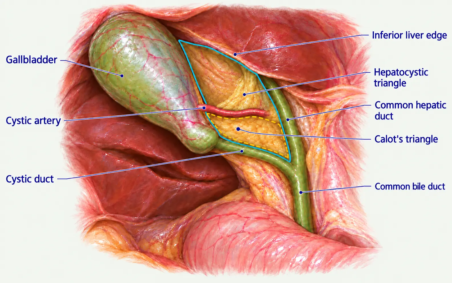
Image source and reuse
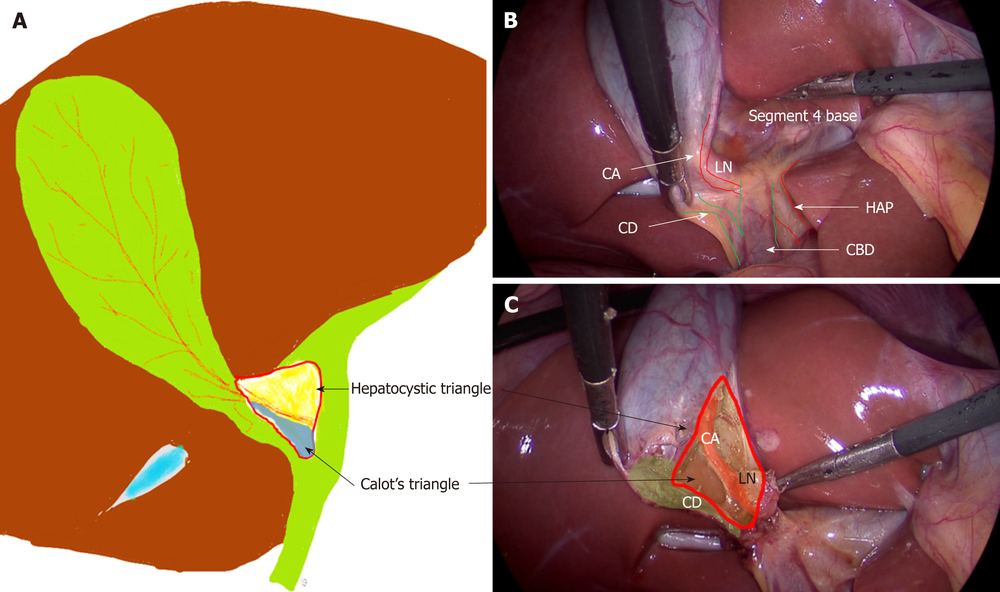
Image source and reuse
The lymph node and what to say in an exam
The cystic lymph node (node of Lund or Mascagni) lies superficial to the cystic artery and marks it; the artery is divided on the gallbladder side of the node (Gupta and Jain 2019). The content that matters most is the one that is not always there: an aberrant right posterior sectoral duct, which joins the common hepatic duct low in about 6 percent of people and the cystic duct in under 1 percent. In either case it crosses the hepatocystic triangle, where it is the structure most often clipped as a “second cystic duct” (Cawich et al. 2021). When examiners say “Calot’s triangle” they usually mean the hepatocystic triangle: give both definitions and say which one the Critical View refers to (Strasberg and Brunt 2010; Gupta and Jain 2019).
03The usual anatomy
- The cystic plate is the fibrous sheet between gallbladder and liver. Stay on the gallbladder side of it; it leads back to the right portal pedicle.
- A cystic duct that has passed a stone can be 5 mm or more. If a medium-large clip will not close it, treat it as the common bile duct.
- Hartmann’s pouch is a stone-induced bulge, not a constant structure, which is why the “infundibular” method of finding the cystic duct is unreliable.
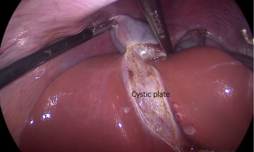
Image source and reuse
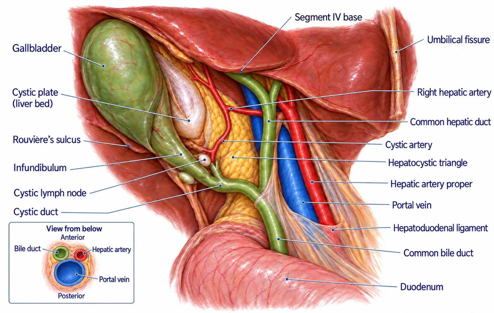
Image source and reuse
More detail and sources
The gallbladder lies in a fossa between segments IV and V and has a fundus, body, infundibulum and neck. The cystic plate is continuous with the hilar plate and, through it, with the sheath of the right portal pedicle. It thickens towards the hilum, and branches of the right pedicle may enter the liver near its lower end. In a small, scarred gallbladder the plate shortens and the liver puckers towards the neck. Dissection deep to the plate then bleeds and, with a fundus-first approach, can enter the right portal pedicle (Gupta and Jain 2019; Gupta 2023). That is the mechanism of the extreme vasculobiliary injuries described under bail-out strategies (Strasberg and Helton 2011).
The cystic duct is usually 2 to 4 cm long and 2 to 3 mm wide, with the spiral valves of Heister. It joins the common hepatic duct at an angle in about 75 percent, runs parallel in about 20 percent and spirals in about 5 percent (Gupta and Jain 2019, citing Abdalla et al. 2013 and Strasberg). Observational The common bile duct is dilated on ultrasound at more than 6 mm in adults with the gallbladder in place and more than 8 mm after cholecystectomy (ASGE 2019).
04Fixed landmarks: B-SAFE, Rouvière’s sulcus, R4U
The “cognitive map” to place before any dissection (Sutherland and Dixon 2017). Tokyo Guidelines 2018 name the sulcus and the base of segment IV as the landmarks to find first (Tokyo Guidelines 2018).
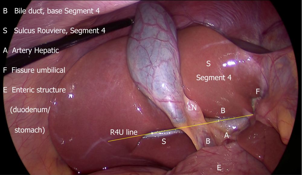
Image source and reuse
- The plane of the sulcus marks the level of the common bile duct: cystic structures lie ventral and cephalad, the main duct dorsal.
- The R4U line runs from the roof of the sulcus across the base of segment IV to the umbilical fissure. Dissect above and lateral to it.
- No sulcus? Extend the line across the base of segment IV to the right, across the hepatoduodenal ligament.
- Say the landmarks out loud before touching the triangle: sulcus, segment IV base, duodenum, duct.
- Start dissection high on the gallbladder, above the line, lateral to the cystic lymph node.
- Treat any tube running below the line, or behind the duodenum, as the common bile duct.
- Rely on the sulcus alone: a quarter of people have none and the closed, slit and scar forms are hard to see.
- Dissect medial to the cystic lymph node or below the line “to clarify”.
- Treat the R4U line as proven: it is a teaching construct from anatomical reasoning and case series.
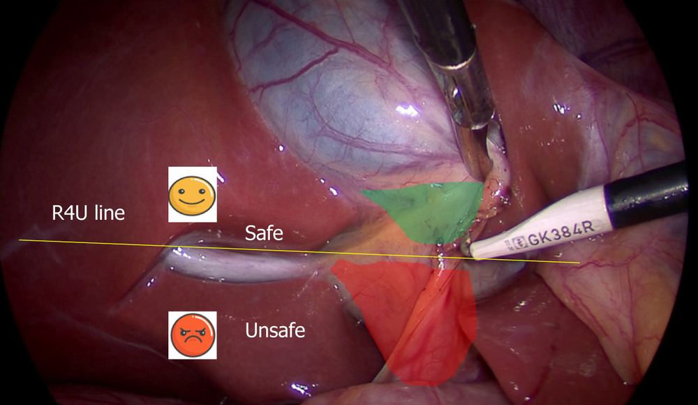
Image source and reuse
More detail and sources
A 2026 systematic review reclassified 57 studies (25,196 people) into one scheme and found a sulcus in 76.9 percent. The types were open 56.7, closed 23.6, slit 14.2 and scar 8.1 percent, with real differences between populations (Cawich et al. 2026). A 2026 Indian series of 100 laparoscopic cholecystectomies found it in 79 percent (Dhanorkar et al. 2026). The sulcus-to-duct relationship was described by Hugh et al. 1997 and is the basis of keeping the posterior dissection cephalad to the sulcus (Gupta and Jain 2019).
Gupta formalised the R4U line and the two zones of the field. The safe zone is above the line and close to the gallbladder. The danger zone, below the line or medial to the cystic lymph node, contains the bile duct and hepatic artery (Gupta and Jain 2019; Gupta 2023). Expert opinion The multi-society guideline endorses the landmarks but does not name the line (Brunt et al. 2020).
05Duct variants that cause injury
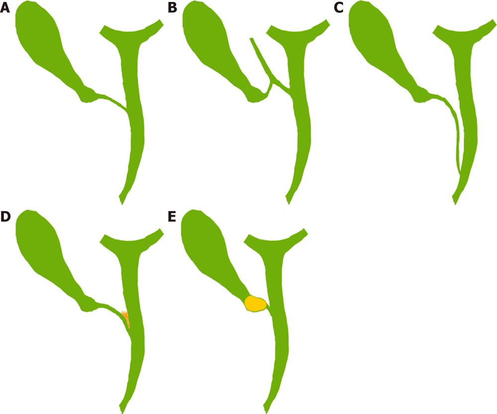
Image source and reuse
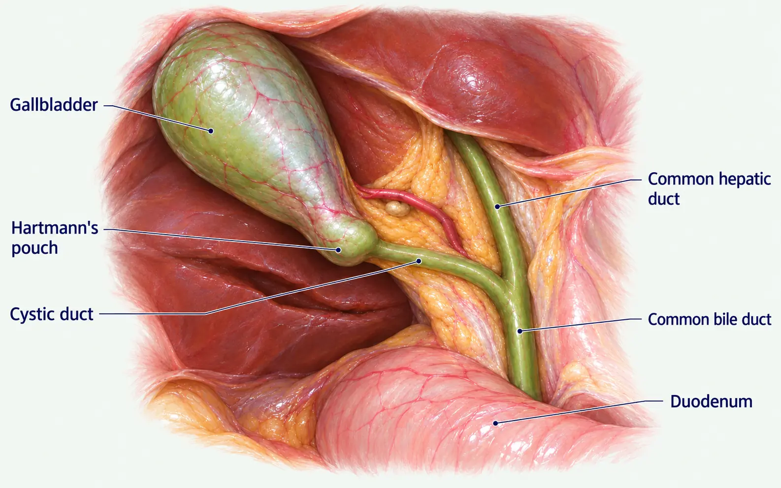
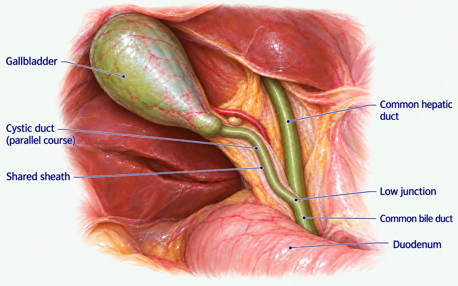
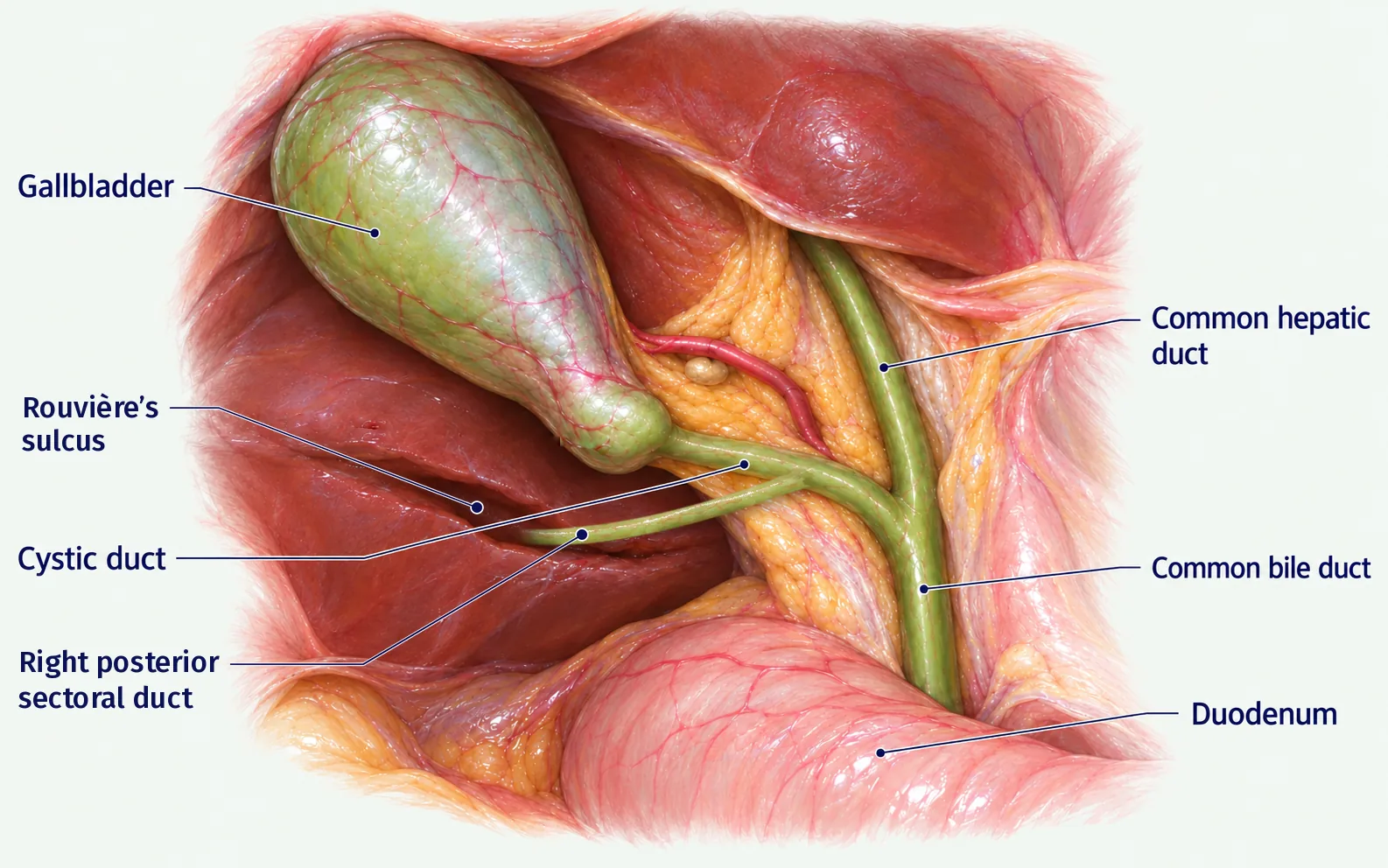
Image source and reuse
More examples (1)
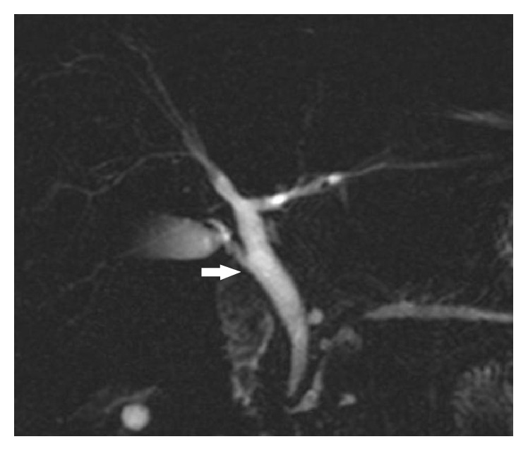
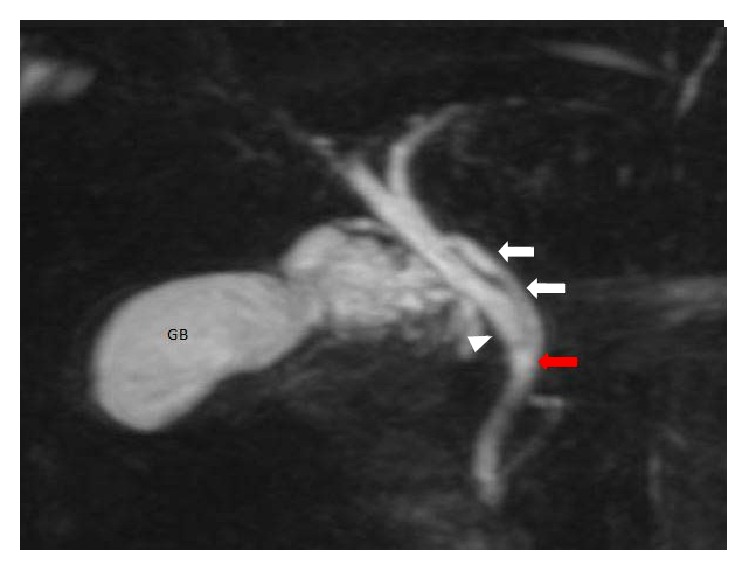
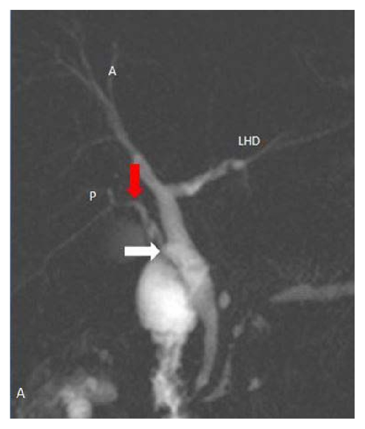
Image source and reuse
Reproduced from Sarawagi R, Sundar S, Gupta SK, Raghuwanshi S. Anatomical variations of cystic ducts in magnetic resonance cholangiopancreatography and clinical implications. Radiology Research and Practice 2016;2016:3021484, Figure 4 (doi:10.1155/2016/3021484), under the CC BY (version not stated by the journal) licence. Reproduced unchanged.
Reproduced from Sarawagi R, Sundar S, Gupta SK, Raghuwanshi S. Anatomical variations of cystic ducts in magnetic resonance cholangiopancreatography and clinical implications. Radiology Research and Practice 2016;2016:3021484, Figure 8 (doi:10.1155/2016/3021484), under the CC BY (version not stated by the journal) licence. Reproduced unchanged.
| Variant | How it fools you | Injury |
|---|---|---|
| Aberrant right posterior sectoral duct joining low, or into the cystic duct | A “second cystic duct” inside the triangle | Strasberg B (occluded) or C (leaking; may be missed on ERCP) |
| Short or effaced cystic duct (impacted stone, Mirizzi) | Traction tents the common duct into the line of the cystic duct | D or E |
| Parallel cystic duct in a common sheath (about 20 percent) | Dissecting “to the junction” strips or burns the CHD; clips narrow it | D, E1, E2 |
| Cystic duct into the right hepatic duct (0.6 to 2.3 percent) | The right hepatic duct is taken for the cystic duct | E4, E5 |
| Subvesical ducts in the liver bed | Divided when dissection goes deep to the cystic plate | A (leak), not misidentification |
- The right posterior sectoral duct drains segments VI and VII. Low, it lies in the triangle and is clipped as a second cystic duct; ERCP may miss the leak if the duct is disconnected.
- This is why SAGES strategy 2 is “understand the potential for aberrant anatomy in all cases”.
Frequencies, how they were measured, and sources
A systematic review pooling 48 imaging and operative studies found the typical confluence in 62.6 percent (study range about 40 to 80 percent) and a triple confluence in 11.5 percent. The right posterior sectoral duct drained into the left duct in 11.5 percent, into the common hepatic duct in 6.4 percent (range about 0.3 to 16) and into the cystic duct in 0.58 percent (up to 3 percent in some series) (Cawich et al. 2021). Subvesical ducts were found in about 4 percent in a systematic review, depending on how they were looked for (Schnelldorfer et al. 2012). Cystic duct course and insertion figures are from Gupta and Jain 2019. Contested Pooled figures come from MR and operative cholangiography, which see every duct; the surgeon dissecting the triangle meets far fewer, and the study-to-study range is wide because of classification and population differences. No reliable population figure exists for a short or absent cystic duct, for a left-sided insertion or for a very long duct; the text says so rather than guessing (SAGES Safe Cholecystectomy Program; Gupta and Jain 2019).
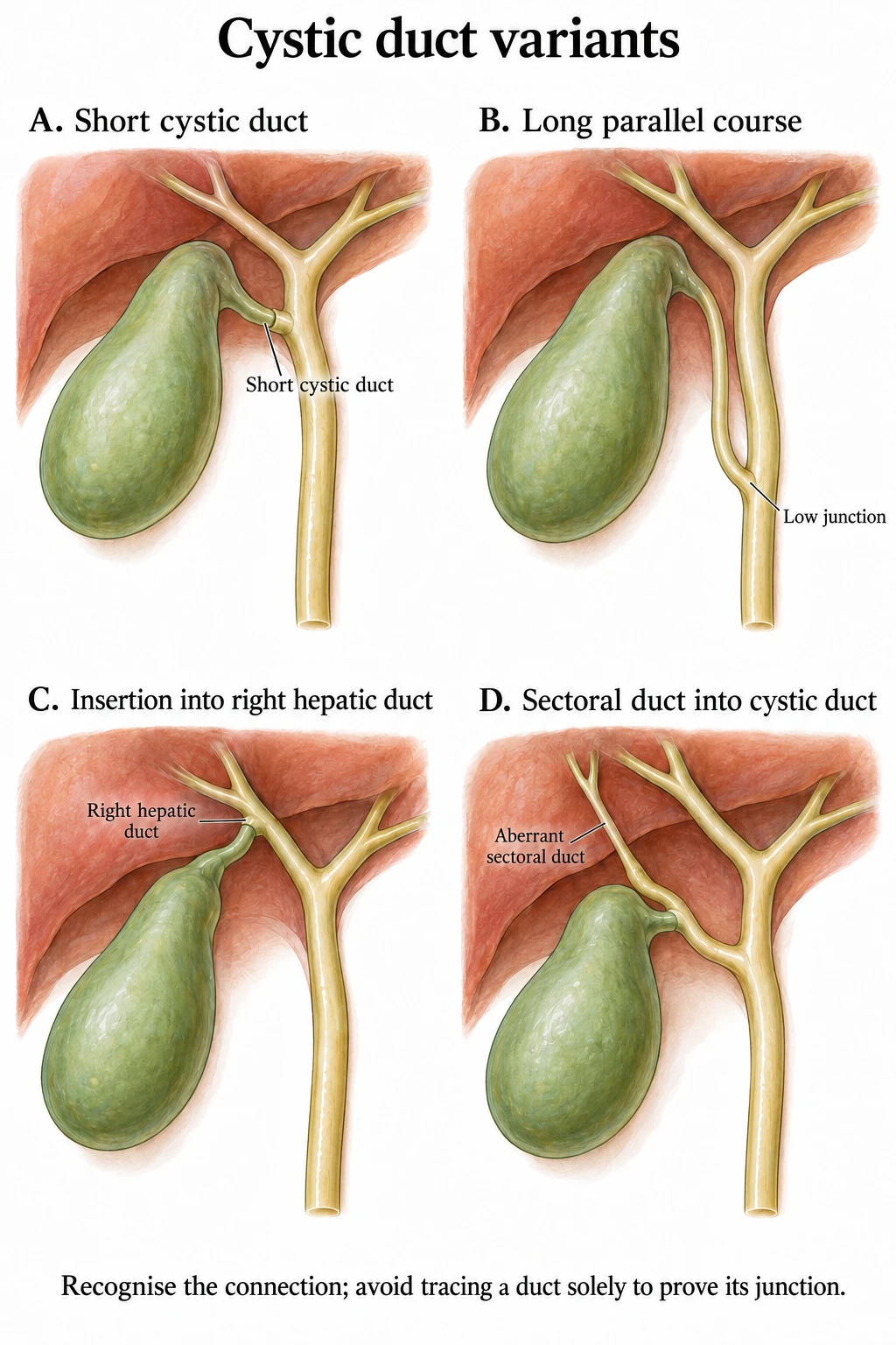
Illustration sources
06Artery variants that cause injury
Pooled data from over 9,800 cases (Andall et al. 2016) and 1,000 transplant donor livers (Hiatt et al. 1994). So one artery in five is not from the right hepatic artery and one in five is not in the triangle.
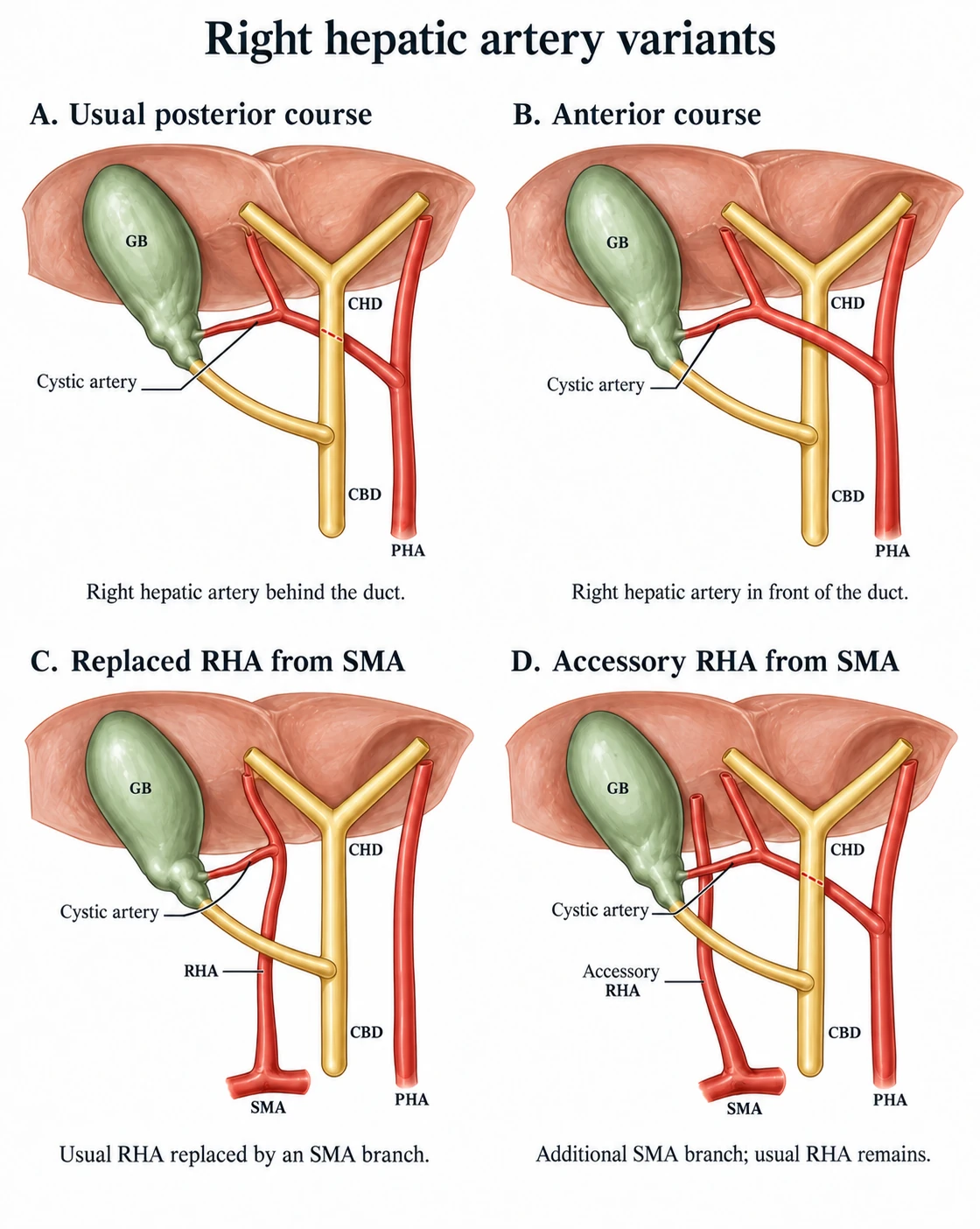
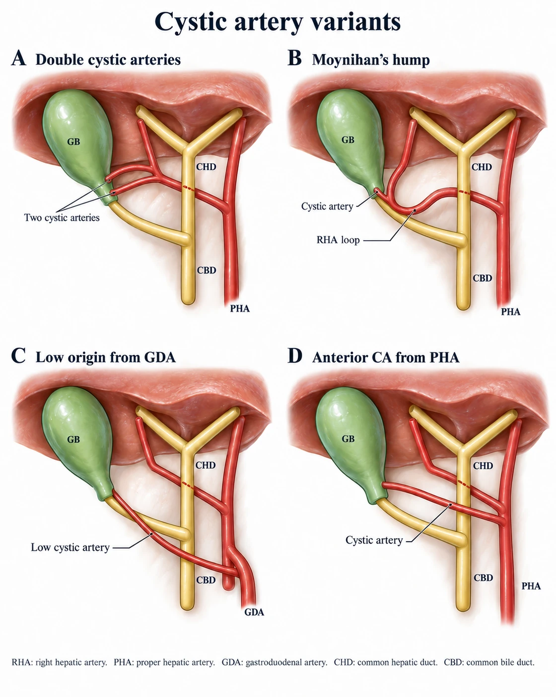
Illustration sources
Moynihan’s hump (caterpillar turn)
Anatomy. The right hepatic artery loops into the triangle against the gallbladder neck and gives a short cystic artery.
Recognise. A wide, tortuous, strongly pulsating “cystic artery” hugging the neck.
Injury. Clipped or torn as the cystic artery: right hepatic artery injury, often with a duct injury.
About 7% of cases (studies 1 to 13%)
Replaced or accessory right hepatic artery
Anatomy. From the superior mesenteric artery, up behind the portal vein and bile duct, then lateral to the duct and close to the cystic duct.
Recognise. A large artery running up beside the cystic duct; pulsation in the lateral ligament where none should be; visible on CT behind the portal vein.
Injury. Hit during posterior or lateral dissection; division devascularises the right liver and the duct.
About 11% of people
Double cystic artery
Anatomy. Separate anterior (superficial) and posterior (deep) arteries; the deep artery may run behind the cystic duct.
Recognise. A second artery after the first is clipped is expected, often seen only in the posterior (doublet) view.
Injury. Bleeding from the unseen deep branch; identify it on the gallbladder side before clipping.
About 9% of people
Cystic artery from the gastroduodenal, left or proper hepatic artery
Anatomy. Does not pass through the triangle. The gastroduodenal variant runs below the cystic duct and in front of the bile duct.
Recognise. Met low and medially, before the triangle is opened, or crossing in front of the common hepatic duct.
Injury. Bleeding before the surgeon has oriented; blind diathermy next to the duct.
Anterior to the CHD in about 18%
- Dissect close to the gallbladder wall, lateral to the cystic lymph node.
- Clip only the short vessel that actually enters the gallbladder wall.
- Confirm two structures from both sides before dividing anything.
- Clip a wide or pulsating artery as “the cystic artery”.
- Apply blind clips or diathermy to pulsatile bleeding in the triangle.
- Chase an artery medially or behind the bile duct.
- A duct injury plus a right hepatic artery injury is a vasculobiliary injury: strictures, abscesses, sometimes hepatectomy.
- The supraduodenal duct is fed by axial vessels at 3 and 9 o’clock, mainly from below. Circumferential dissection or diathermy near the duct causes late ischaemic strictures.
Prevalence data in full, with sources
Cystic artery. Origin from the right hepatic artery 79.0 percent; course through the triangle 81.5 percent (5,427 of 6,661); anterior to the common hepatic duct 17.9 percent (485 of 2,704); anterior to the common bile duct 5.4 percent (228 of 4,202); short artery 9.5 percent (98 of 1,037); multiple arteries 8.9 percent; inferior to the cystic duct 4.9 percent (38 of 770) (Andall et al. 2016). The right hepatic artery passes behind the common hepatic duct in about 87 percent (Gupta and Jain 2019, citing Skandalakis). A 2025 systematic review of 22 studies reported a non-standard origin in nearly two thirds of the studies. A double artery was reported in more than half, and an anterior course in about a third (Schiewe et al. 2025). When the cystic artery arises from the gastroduodenal or left hepatic artery it does not pass through the triangle and cannot be found by dissecting there (Gupta and Jain 2019).
Moynihan’s hump. Systematic review of 16 studies: 6.9 percent of 1,077 cases (operative about 7 percent, cadaver 6.8 percent, individual studies about 3 to 13 percent), and 1.3 percent in the authors’ own 230 cases. The loop passed behind the common hepatic duct in 60 percent and in front of it in 40 percent. It was single in 55 percent and double in 45 percent (Marano et al. 2018). Contested The spread reflects how hard the variant is to define and to notice. Expert opinion There is no validated size threshold. The rule on this site: any artery that looks wider than a normal cystic artery, or pulsates visibly, is the right hepatic artery until proven otherwise (Marano et al. 2018; Gupta and Jain 2019).
Replaced and accessory right hepatic artery. 10.6 percent (106 of 1,000 donor livers), with normal anatomy in 75.7 percent (Hiatt et al. 1994); 8.7 to 11.0 percent replaced and 7.0 to 10.7 percent accessory across imaging and operative series, normal anatomy 55 to 81 percent (Samuolyte et al. 2025). Course and recognition from Gupta and Jain 2019.
Double cystic artery and the posterior view. The deep branch and its junction with the anterior branch may only be visible when the gallbladder is flipped to the left (Gupta 2023).
Vasculobiliary injury. 39 percent (19 of 48) of referred bile duct injuries had a concomitant vascular injury on CT angiography, the right hepatic artery most often; earlier series 12 to 61 percent (Singla et al. 2025); consequences from Strasberg and Helton 2011. Contested The range depends on whether angiography was routine and on referral bias.
Blood supply of the bile duct. Axial vessels at 3 and 9 o’clock, fed mainly from below (retroduodenal and gastroduodenal arteries) and to a lesser extent from above (right hepatic artery) (Northover and Terblanche 1979).
07Gallbladder variants and other traps
- Intrahepatic gallbladder: no plane to the cystic plate; expect liver-bed bleeding (Parkland grade 4).
- Left-sided gallbladder: rare; the cystic duct often crosses in front of the common duct; mirror the ports.
- Duplication, agenesis, phrygian cap: usually radiological curiosities, but a gallbladder that cannot be found at laparoscopy is a stop sign, not an invitation to explore: image (MRCP) rather than dissect, because agenesis is real and the ducts lie where you would be looking. A missed second gallbladder is a cause of recurrent symptoms.
- Mirizzi syndrome: the pathological short cystic duct; the triangle is obliterated. See special situations.
08What examiners ask
- Define both triangles and say which one the Critical View of Safety uses.
- Name the structure in Rouvière’s sulcus (right posterior sectoral pedicle) and what the sulcus tells you (the level of the CBD).
- Describe the blood supply of the bile duct and why diathermy near it causes late strictures.
- Explain why an aberrant sectoral duct leak may not show on ERCP.
- Quote the cystic artery frequencies (79, 81, 9, 10 percent) and describe Moynihan’s hump and the replaced right hepatic artery: anatomy, recognition, injury.
Test yourself further: Practise questions on Anatomy and variants in the Exam zone.