Quick reference
Safety checklist, decision pathways and key figures.
Designed and developed by Dr Prasad Bhukebag
01Safety checklist
Before the list
- Indication confirmed; imaging reviewed personally: duct diameter, stones in duct, liver tests, any endoscopic retrograde cholangiopancreatography (ERCP) done.
- Difficulty predicted (age, male, acute cholecystitis, thick wall, contracted gallbladder, previous cholecystostomy, ERCP, cirrhosis); operation listed with the right surgeon and the right time of day.
- Consent includes bile duct injury with a figure, conversion, subtotal cholecystectomy, leak, persisting symptoms.
- Antibiotic prophylaxis for acute cholecystitis and risk groups; none routinely for simple elective cases.
- Cholangiography equipment available and staff able to set it up; indocyanine green (ICG) given 30 minutes or more before incision if it is to be used.*
Time-out 1 (before incision)
- WHO checklist items; plus: imaging result stated, difficulty grade expected, cholangiography plan, bail-out plan named, senior help identified.
Entry and exposure
- Safe entry (open at the umbilicus, or optical); previous surgery means away from the scar.
- Grade the gallbladder at first sight (Parkland or Nassar), say it out loud, record it.
- Decompress a tense gallbladder before grasping it. Retract the fundus towards the right shoulder and the infundibulum laterally to open the triangle.
- Find Rouvière’s sulcus and the base of segment IV; stay above the R4U line. Identify the B-SAFE landmarks.
Dissection
- Dissect on the gallbladder, starting at the infundibulum, posterior window first, then anterior, and clear the hepatocystic triangle of fat and fibrous tissue.
- Separate the lower third of the gallbladder from the cystic plate.
- Do not clip or divide the cystic duct or artery until all three CVS criteria are met and seen from the front and the back (doublet view). During dissection, use energy sparingly on tissue lifted away from the bile ducts.
- Use intra-operative biliary imaging, particularly X-ray cholangiography, when anatomy is uncertain or bile duct injury is suspected. For suspected duct stones, use cholangiography or laparoscopic ultrasound; ICG fluorescence does not assess the duct lumen or exclude stones. ICG is an adjunct to anatomical identification, not a substitute for a safe dissection.
Time-out 2 (before clipping)*
- Say the three criteria out loud; ask the assistant to agree; photograph or record the view.
- Count the structures: two, entering the gallbladder, and nothing else.
- Clip and divide the cystic duct and artery close to the gallbladder; never clip a structure you cannot see all of.
If the CVS cannot be achieved: the stop rule
- Stop. No further dissection in the hepatoduodenal ligament. Tell the team out loud that you have stopped.
- Call for a second surgeon. Image the ducts.
- Choose a bail-out: subtotal cholecystectomy (fenestrating or reconstituting), fundus-first only if the cystic plate is safe, conversion only if it helps, or abandon and drain. Record the reason.
Completion and sign-out
- Inspect the liver bed and the clips at low pressure; wash out; retrieve spilled stones and document any left; drain selectively.
- Close 10 mm port fascia under vision.
- Sign-out: CVS achieved (yes/no and how); any concern about the duct; leak, spillage, bleeding; plan and who to call.
- Operation note written the same day with the criteria, the grade, the imaging and the decisions.
If a bile duct injury is suspected or found
- Stop. Do not clip, suture or attempt repair unless you are a hepatobiliary surgeon.
- Define the anatomy by cholangiography if possible; photograph.
- Drain the area widely; close; tell the patient; refer the same day to a hepatobiliary unit.
Checklist sources and qualifications
Print this. It condenses the SAGES six strategies, the 2020 multi-society consensus, Tokyo 2018 and the WSES guidance into the steps of one operation (SAGES Safe Cholecystectomy Program; Brunt et al., Ann Surg 2020). Guideline This is a practical checklist combining guidelines and expert practice, not a list of mandatory evidence-based requirements. In particular, CVS documentation is useful for review but was not mandated by the 2020 panel; bail-outs are chosen by circumstances, not a fixed sequence. Recommendation strength and certainty are available on the safety page.
02The difficult gallbladder
Orientate and assess difficulty
Identify fixed landmarks; optimise exposure, decompress if needed and assess progress.
Can all three CVS criteria be achieved safely?
Yes
Confirm CVS, pause, document
Use anterior and posterior views. Once identification is secure, clip, divide and complete.
No / progress stalls
STOP and obtain help
No clips on an uncertain structure. Ask an experienced colleague; use IOC when anatomy is uncertain or injury is suspected. ICG is an adjunct.
After STOP and reassessment / imaging, is the anatomy still undefined?
No
Choose an approach with a safe plane
Fundus-first total dissection is considered only when anatomy is defined and the plane is safe. Stop if the plane is lost.
Yes
Prefer subtotal over total fundus-first
When CVS cannot be obtained and anatomy remains undefined, consider subtotal cholecystectomy if it can be performed safely.
Conversion is one safety option
Convert when it improves access or control. Conversion alone does not resolve distorted anatomy.
Still unsafe? Stop or abort
Use drainage or cholecystostomy when appropriate; control infection and arrange specialist care. Document the reason and explain it to the patient.
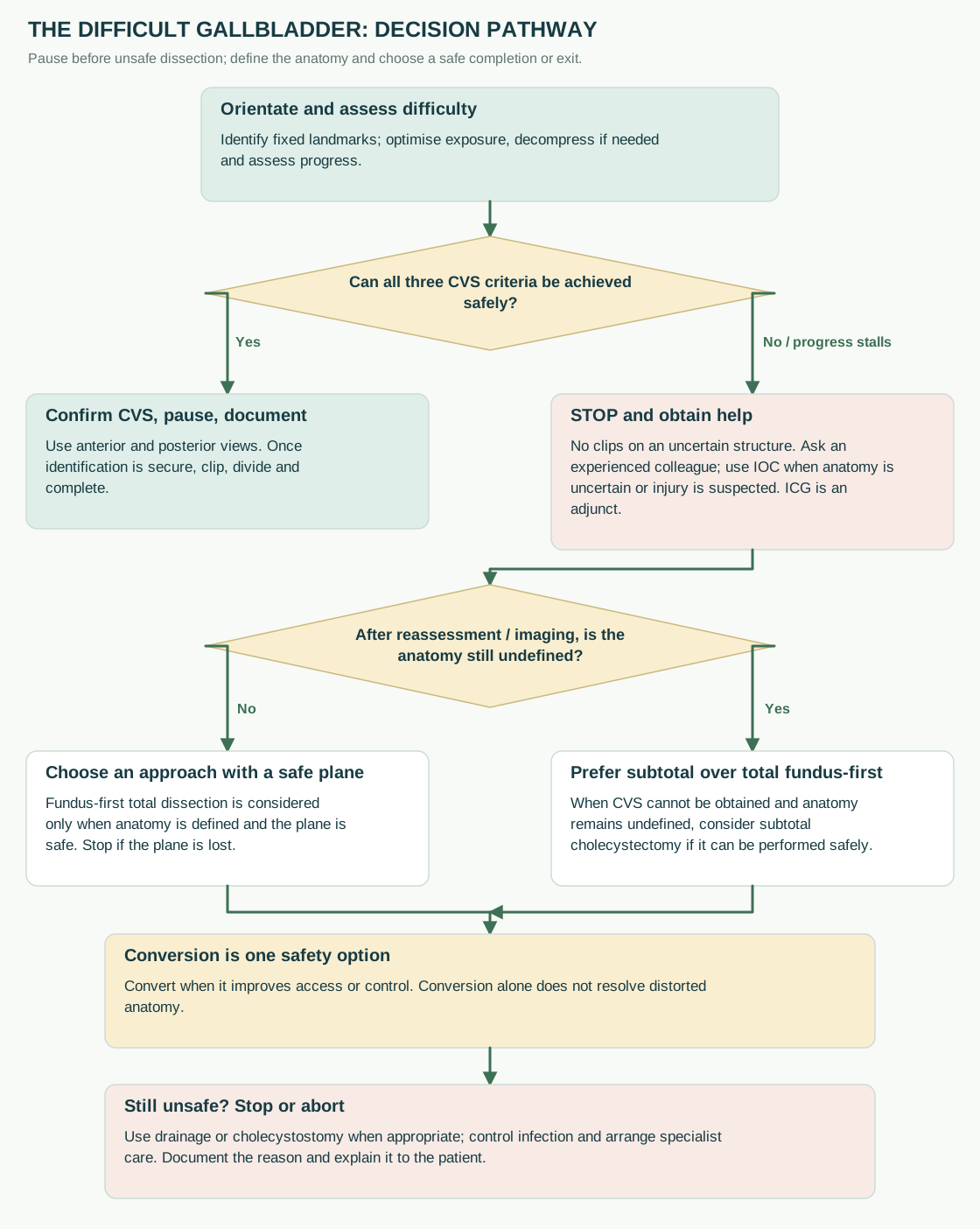
Diagram sources
03Suspected bile duct injury
Choose the presentation
Recognised during surgery
Stop clipping and energy use. Avoid further hilar dissection. Call an experienced HPB surgeon. Cholangiography only if safe. Without appropriate expertise: control leakage with drainage and arrange transfer. Selected immediate repair requires specialist judgement.
Suspected after surgery
Resuscitate; assess blood tests and treat sepsis. CT: collections and vascular perfusion. Drain collections when indicated. MRCP maps duct anatomy. Select ERCP or percutaneous cholangiography according to continuity and access.
For either presentation
Define continuity, injury level and vascular involvement
Agree the next step with the hepatobiliary team.
Choose the injury branch
A / selected small D · Leak with main-duct continuity
Drainage and endoscopic treatment may be appropriate after assessment.
B / C · Sectoral duct injury
Obstructed or disconnected territory. ERCP may miss a disconnected duct; HPB assessment and tailored imaging / drainage are required.
E / large D / vascular · Major or complex injury
Prompt referral. Control sepsis and leakage, support nutrition and map the injury. Definitive reconstruction is planned by the HPB team.
Timing and communication
SAGES–AHPBA 2025 conditionally favours repair after 6 weeks (very low certainty); selected earlier repair requires expertise. Explain the injury, preserve the operative record and video, and send the imaging with the referral.
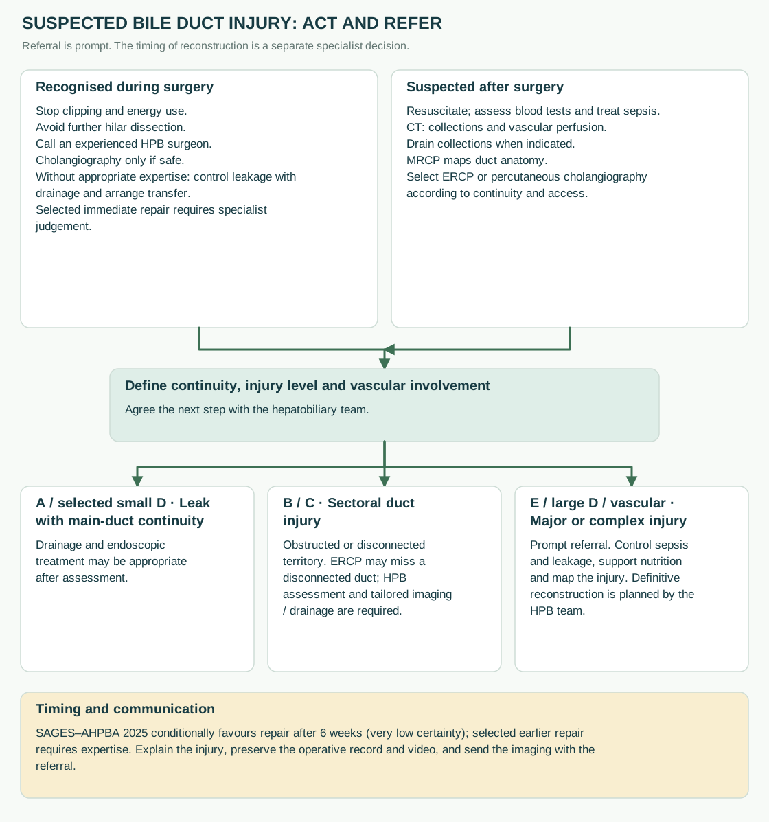
Diagram sources
04Numbers to remember
| Measure | Figure | Source |
|---|---|---|
| Major bile duct injury, laparoscopic cholecystectomy | 0.2 to 0.4 percent (about 1 in 300 to 500) | Pucher 2018; AMBROSE 2025; Neel 2026 |
| Bile leak of any kind | About 1 percent | AMBROSE 2025 |
| 30-day mortality, all cholecystectomies | 0.1 to 0.4 percent; much higher over 80 and in emergency | Pucher 2018; AMBROSE 2025; Kamarajah 2020 |
| Early LC in acute cholecystitis: window | Within 7 days of admission and 10 days of symptom onset (WSES); within 1 week of diagnosis (NICE); “as soon as possible” if fit (Tokyo 2018) | Pisano 2020; NICE QS104; Okamoto 2018 |
| Early vs delayed LC, acute cholecystitis | No difference in bile duct injury; 4 days shorter total stay; about 18 percent of delayed group need urgent surgery while waiting | Gurusamy Cochrane 2013 |
| High-risk acute cholecystitis: LC vs cholecystostomy | Major complications 12 vs 65 percent favouring LC | CHOCOLATE, BMJ 2018 |
| Antibiotic prophylaxis omitted in mild to moderate acute cholecystitis | Infectious complications 12.6 vs 7.1 percent; omission not recommended | PEANUTS II, BJS 2022 |
| ICG dose and timing | 2.5 mg IV, 30 minutes or more before incision (FALCON); 0.05 mg/kg weight-based (Lehrskov); giving the dye more than 3 hours before gives a better duct-to-liver picture | FALCON 2023; Lehrskov 2020; DOTIG 2025; Boogerd 2017 |
| ICG and bile duct injury (observational) | 0.25 vs 0.40 percent with vs without ICG; relative risk 0.62 | Neel 2026 |
| Fenestrating vs reconstituting subtotal: bile leak | Higher with fenestrating (odds ratio about 0.29 favouring reconstituting; 22 vs 7 percent in a prospective series) | Motter 2024; Johnson 2026 |
| Bile duct injury: referral and repair timing | Refer promptly to an experienced hepatobiliary team. The team determines reconstruction timing: SAGES–AHPBA 2025 conditionally favours delayed repair beyond 6 weeks, while early repair may be considered in selected patients. Control sepsis and drain bile as needed while planning definitive treatment; hepaticojejunostomy is the established reconstruction for major injuries | SAGES–AHPBA 2025; WSES 2021; Schreuder 2020 |
| Incidental gallbladder cancer re-resection | Fit patients with resectable T1b–T3 and no metastases: hepatobiliary assessment for re-resection; the 4–8-week survival finding is observational | AHPBA consensus 2015; Ethun 2017 (observational) |
| Bile duct injury, robotic vs laparoscopic (US Medicare, observational) | 0.7 vs 0.2 percent (2010 to 2019); 0.38 vs 0.18 percent, relative risk 2.09 (2020 to 2023) | Kalata 2023; Mullens 2026 |
| Single-incision vs four-port: hernia at 1 year | 8.4 vs 1.2 percent | Marks 2013 |
| Bile duct exploration: transcystic vs choledochotomy, bile leak | 1.5 vs 8.8 percent; retained stones 4.4 percent overall; 30-day mortality 0.4 percent | R-ALiCE 2026 |
| T-tube vs primary closure after choledochotomy | Routine T-tube: longer stay and operating time, without demonstrated benefit in the reviewed trials; use drainage selectively | Gurusamy Cochrane 2013 |
| Gallbladder polyps | 10 mm or more: operate; 6 to 9 mm with a risk factor: operate; if fit and accepting surgery. Surveillance: 6–9 mm without risk factors or ≤5 mm with a risk factor, at 6, 12 and 24 months; ≤5 mm without risk factors needs no follow-up | Foley 2022 |
| Learning curve | 90 percent of early injuries within a surgeon’s first 30 cases; modelled risk 1.7 percent at case 1, 0.17 percent at case 50 | Moore and Bennett 1995 |
| Litigation | England: 65 percent of 418 claims succeeded, mean £102,827. USA: 40 percent of 745 claims paid, median $264,650; bile duct injury recognised intra-operatively in 19 percent | Alkhaffaf 2010; Gartland 2019 |
05Glossary
- AAST grade
- American Association for the Surgery of Trauma anatomical severity grade for acute cholecystitis: I uncomplicated; II empyema, gangrene or emphysema; III perforation with local contamination; IV pericholecystic abscess or fistula; V perforation with generalised peritonitis.
- ASA grade
- American Society of Anesthesiologists physical status, I (healthy) to V (moribund).
- B-SAFE
- Mnemonic for fixed landmarks: Bile duct, Sulcus of Rouvière, hepatic Artery, umbilical Fissure, Enteric (duodenum).
- Bail-out
- A planned alternative to total cholecystectomy when the CVS cannot be achieved safely: subtotal cholecystectomy, fundus-first, conversion or abandonment.
- BDI
- Bile duct injury.
- Bismuth classification
- Classification of bile duct strictures by their height relative to the hilum, I to V.
- Calot’s triangle
- As originally described, the triangle between the cystic duct, the common hepatic duct and the cystic artery; now usually used loosely for the hepatocystic triangle.
- CBD, CHD
- Common bile duct; common hepatic duct.
- Cholecystostomy (PTGBD)
- Percutaneous transhepatic gallbladder drainage: a tube placed into the gallbladder under imaging guidance.
- Clavien-Dindo
- Grading of surgical complications by the treatment they need, I to V.
- Conversion
- Change from laparoscopic to open operation during the procedure.
- CVS
- Critical View of Safety: the three criteria that identify the cystic duct and artery before they are divided.
- Cystic plate
- The fibrous sheet between the gallbladder and the liver, continuous with the hilar plate; the plane of dissection stays on the gallbladder side of it.
- Doublet view
- The CVS seen and documented from both the anterior and the posterior aspect.
- Duct of Luschka
- A subvesical bile duct in the cystic plate or liver bed; a source of post-operative bile leak.
- ERCP
- Endoscopic retrograde cholangiopancreatography.
- EUS
- Endoscopic ultrasound.
- Fenestrating subtotal cholecystectomy
- Subtotal cholecystectomy in which the gallbladder remnant is left open and the cystic duct is closed from inside or left to drain.
- Fundus-first (top-down, retrograde)
- Dissection of the gallbladder off the liver from the fundus towards the neck.
- G10 score
- Ten-item intra-operative difficulty score (Sugrue).
- Hannover classification
- Classification of bile duct injury that adds vascular involvement and the level of injury (A to E with subtypes).
- Hartmann’s pouch
- A bulge of the infundibulum or neck produced by a stone or chronic disease; not a constant structure, so it is an unreliable guide to the cystic duct.
- Hepaticojejunostomy (HJ)
- Anastomosis of the hepatic duct (or ducts) to a Roux loop of jejunum; the standard repair for major bile duct injury.
- Hepatocystic triangle
- Triangle bounded by the cystic duct, the common hepatic duct and the inferior edge of the liver; the area cleared to obtain the CVS.
- ICG
- Indocyanine green, a dye excreted in bile that fluoresces under near-infrared light.
- Infundibulum
- The funnel-shaped part of the gallbladder between the body and the neck, leading into the cystic duct.
- Infundibular technique
- Identification of the cystic duct by the flare where it meets the infundibulum; unreliable in inflammation and a cause of injury.
- IOC
- Intra-operative cholangiography with X-ray contrast.
- LUS
- Laparoscopic ultrasound.
- Mirizzi syndrome
- Obstruction of the common hepatic duct by a stone impacted in the gallbladder neck or cystic duct, with or without a fistula into the duct.
- MRCP
- Magnetic resonance cholangiopancreatography.
- Nassar grade
- Intra-operative difficulty grade 1 to 5 based on gallbladder, cystic pedicle and adhesions.
- Parkland grade
- Intra-operative difficulty grade 1 to 5 based on the appearance of the gallbladder at first view.
- Reconstituting subtotal cholecystectomy
- Subtotal cholecystectomy in which the remnant is closed to form a small pouch.
- Rouvière’s sulcus
- A cleft on the inferior surface of the right liver lobe that marks the level of the right posterior pedicle; dissection stays above it.
- R4U line
- Imaginary line from Rouvière’s sulcus to the base of segment IV and the umbilical fissure; the safe zone of dissection is above it.
- Stewart-Way classification
- Mechanistic classification of bile duct injury, classes I to IV.
- Strasberg classification
- Classification of bile duct injury, A to E (E1 to E5 by level), the standard in the literature.
- Tokyo Guidelines 2018 (TG18)
- Diagnostic criteria and severity grading (I to III) of acute cholecystitis and cholangitis, with flowcharts for management.
- Vasculobiliary injury
- A bile duct injury combined with injury to the right hepatic artery or portal vein.
- Zone of risk
- The hepatoduodenal ligament below the R4U line and medial to the gallbladder neck, where the bile duct and hepatic artery lie.
Review information and key reminders
Reviewed against its sources on 3 November 2026. Latest source: Multi-society safe cholecystectomy consensus, Ann Surg 2020. Full page: about 9 min read. How evidence is marked.
- Before clipping: the three Critical View criteria, from both sides, said out loud.
- When the view will not come: stop, get help, image, bail out.
- When an injury is suspected: stop, drain, refer. Do not repair.
This is a teaching reference. Use it alongside local protocols and clinical judgement.