Bile duct injury
Bile duct injury is uncommon, but when it happens the first decisions decide whether the patient has one operation or a lifetime of them.
Reviewed against its sources on 29 October 2026. Open checks: the share of major injuries with a right hepatic artery injury varies widely between referral series and is given only as a rough proportion.
- Classify by Strasberg (A to E) and say whether an artery is injured (Hannover); the class describes, it does not prescribe.
- On the table: stop, no blind clips, no repair without a hepatobiliary surgeon, drain, refer the same day.
- Bile, pain, fever or jaundice in the first days is a leak or obstruction until proved otherwise: image, drain, refer early.
01How common, and how serious
- Higher in acute cholecystitis, men, the elderly and difficult operations.
- The consequences are out of proportion to the frequency: higher long-term mortality, multiple procedures, lost quality of life.
- In the United Kingdom bile duct injury is the commonest and most successful cause of litigation after laparoscopic cholecystectomy.
Sources
Pooled analysis: bile duct injury 0.32 to 0.52 percent, falling from 0.69 percent in the 1990s to 0.22 percent in 2010 to 2015 (Pucher et al., Surg Endosc 2018). AMBROSE: bile leak 1.3 percent and severe injury (Strasberg B to E) 0.2 percent (AMBROSE Collaborative, Ann Surg 2025). Estonian population study: 0.81 percent overall including minor injuries, 0.13 percent major, no decline over 11 years, mortality 4.6 percent among injured patients (Reinsoo et al., Eur J Trauma Emerg Surg 2023). Litigation: Alkhaffaf and Decadt, Ann Surg 2010.
02Classification
Strasberg (1995): the one every guideline uses
| Type | Injury | Presents as | Treatment |
|---|---|---|---|
| A | Leak from a minor duct in continuity: cystic stump or subvesical (Luschka) duct | Bile in the drain or a collection within days | Drain; endoscopic retrograde cholangiopancreatography (ERCP) with sphincterotomy or stent |
| B | Occlusion of part of the tree, almost always an aberrant right posterior sectoral duct | May present late with pain, cholangitis or segmental atrophy | HPB assessment; management depends on symptoms, obstruction and the liver territory involved |
| C | Transection of an aberrant right sectoral duct, unligated | A leak that may be missed on ERCP because the duct is disconnected | Drain; PTC or magnetic resonance cholangiopancreatography (MRCP) to find it; ligation, anastomosis or resection |
| D | Lateral injury of a major duct, continuity kept: partial clip, burn, tear | Leak, or late stricture if thermal | Selected small injuries: endoscopic treatment after specialist assessment; large or thermal injuries: HPB referral |
| E1 | Circumferential, more than 2 cm below the confluence (Bismuth I) | Leak and biloma, or peritonitis without a drain; jaundice if clipped; cholangitis; late stricture | Roux-en-Y hepaticojejunostomy by a hepatobiliary surgeon at the right time; E4 and E5 may need separate anastomoses or resection |
| E2 | Less than 2 cm below the confluence (II) | ||
| E3 | At the confluence, right and left still joined (III) | ||
| E4 | Confluence destroyed, right and left separated (IV) | ||
| E5 | Aberrant right duct plus common hepatic duct (V) |
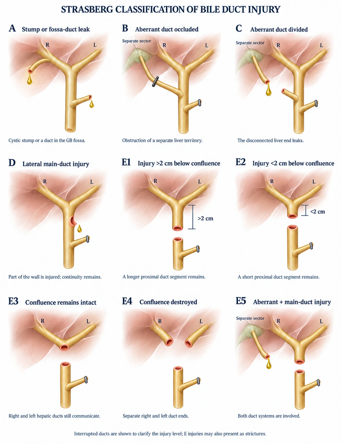
Illustration sources
Bismuth (1982)
Strictures graded by the healthy duct left below the confluence: I over 2 cm; II under 2 cm; III hilar, confluence intact; IV confluence separated; V aberrant right duct involved. E1 to E5 map onto it.
Prognostic: higher type, harder repair, more restricture
Hannover (2007)
A peripheral leak; B occlusion; C tangential injury; D transection; E late stricture, each subgraded by size and level; a suffix names any injured vessel (“D2 d”).
Predicts the operation needed
Stewart-Way
By mechanism. I common duct mistaken but recognised before division; II lateral damage from clips or cautery; III common duct transected, usually with a segment excised (the classic injury, the commonest); IV right hepatic duct or artery mistaken.
ATOM (EAES 2013)
Records Anatomy, Time of detection and Mechanism. WSES recommends it for reporting because it captures what the others leave out.
More detail and sources
Strasberg: Strasberg et al., J Am Coll Surg 1995. Hannover in full: A peripheral leak (A1 cystic duct; A2 gallbladder bed); B occlusion of the main duct without wall injury (B1 incomplete; B2 complete); C tangential injury (C1 under 5 mm; C2 over 5 mm below the confluence; C3 at the confluence; C4 above it); D complete transection (D1 no defect, below the confluence; D2 with loss of length, below; D3 at; D4 above); E late stricture (E1 under 5 mm; E2 over 5 mm; E3 at; E4 above the confluence); vascular injury as a suffix (Bektas et al., Br J Surg 2007). Stewart-Way: Way et al., Ann Surg 2003. ATOM: anatomy (main or non-main duct, occlusion or division, partial or complete, with or without vascular injury), time (intra-operative, early or late post-operative) and mechanism (mechanical or energy) (Fingerhut et al., Surg Endosc 2013). Guideline
03Mechanisms
| Mechanism | How | Result |
|---|---|---|
| Misidentification | The common or right hepatic duct is followed upward “until it enters the gallbladder” and excised between clips, often with the right hepatic artery | The classic injury (perceptual illusion) |
| Thermal | Diathermy on or near the duct, or conducted along clips | Late stricture or delayed leak when the burnt wall sloughs |
| Tenting | Cephalad traction tents the common duct into the line of the cystic duct; clips across the junction, or clips on a short cystic duct narrowing the common duct | Partial occlusion, stricture |
| Slipped or migrated clips | On a wide, inflamed cystic duct | Leak |
| Deep to the cystic plate | In a fibrotic bed | Subvesical duct leak, or entry into the right portal pedicle |
| Bleeding control | Blind clips or diathermy in the triangle | Combined ductal and arterial injury |
Sources
Gupta and Jain, World J Gastrointest Surg 2019, citing the Delphi consensus of Iwashita et al., J Hepatobiliary Pancreat Sci 2017.
- The injury is mostly a failure of perception, not of skill, so prevention is a method rather than a talent.
- The method, in order: the Critical View of Safety before anything is clipped or cut; a difficulty grade before the dissection starts; imaging when the anatomy is unclear; a stopping rule for the view that will not come; and a unit culture in which a bail-out is normal.
- Read those five pages in that order as the prevention chapter of this one.
04Recognising the injury on the table
Only about a quarter of injuries are recognised during the operation (252-case human-factors series).
- Bile in the field from the hepatoduodenal ligament, or from a divided structure, with the gallbladder intact.
- A second “cystic duct”, or a structure that goes on beyond where the cystic duct should end.
- A duct bigger than expected, or a “cystic duct” that turns out to run up to the hilum.
- Unexplained bleeding in the triangle: the right hepatic artery, and a duct injury next to it.
- A cholangiogram that does not fill the proximal ducts, or shows extravasation.
Suspect an injury and have not done a cholangiogram? Do one now, through the gallbladder or the duct that has already been opened; converting to do it is justified. Do not cut into or cannulate a further structure you cannot name to get the picture. The 2020 consensus makes a strong recommendation for imaging whenever an injury is suspected. Guideline
Source
- This is Stewart-Way class I, and it is the one injury the operating surgeon can usually put right. It is recognised when the “cystic duct” proves too long, too wide or heads for the hilum, or when the cholangiogram shows the catheter in the common duct.
- Take the clip off with a clip remover; do not cut it away, and do not dissect further to “check”.
- Image through the cystic duct: the whole tree should fill freely and nothing should leak. Then find the real cystic duct from the gallbladder downwards.
- Look at the wall. A removed clip leaves a crush mark. If the wall is bruised or torn, or has had any contact with diathermy, drain the area and telephone the hepatobiliary unit from theatre. Never suture a burnt wall: it sloughs or strictures weeks later.
- Write it down and tell the patient, even when the cholangiogram is normal, and arrange liver tests at follow-up. A crush or thermal injury can present as a stricture months later, and an undocumented near miss is indefensible then.
Expert opinion Drawn from the mechanism analysis of Way et al. 2003 and the management principles of the guidelines cited on this page; no guideline addresses the near miss directly.
05What to do and what not to do immediately
Do not attempt to repair a major bile duct injury unless you are a hepatobiliary surgeon with experience of biliary reconstruction.
- Stop. No clips, no diathermy, no tidying up. Tell the team.
- Call for help: the most experienced surgeon in the building, and the hepatobiliary unit by telephone from theatre.
- Define the injury: cholangiography through the divided duct or the gallbladder; note whether the right hepatic artery pulsates.
- Not a hepatobiliary surgeon? Large closed-suction drain to the hilum; leave the duct ends alone; a controlled external fistula is the aim; close and transfer. The one exception is a small, sharp, non-thermal lateral injury with the duct otherwise intact (a Strasberg D from a scissor nick or a partial clip): after cholangiography and a telephone call to the hepatobiliary unit, an experienced general surgeon may close it with fine absorbable sutures over a T-tube or a stent and leave a drain. Everything else, and every thermal injury, is drained and referred. Expert opinion
- Hepatobiliary surgeon present, patient stable, no sepsis, clean transection, no arterial injury: immediate Roux-en-Y hepaticojejunostomy is reasonable. Complex and vasculobiliary injuries: drain and plan, even in expert hands (WSES).
- Tell the patient and family when awake, in plain words. The duty of candour applies.
- Document: drawing, clips, drain, images, video. Report the incident.
- Attempt an end-to-end anastomosis or a hepaticojejunostomy if you do not do them regularly.
- Place more clips, or look for “the other end” with diathermy.
- Ligate the proximal duct to “stop the leak”: it obstructs the whole liver and makes repair harder.
- Leave the patient without a drain and hope.
- Delay the call because the patient “looks fine”: bile peritonitis is painless for hours.
Referral and repair: guidance and qualifications
The 2020 consensus makes a strong recommendation for prompt referral to an experienced surgeon or hepatobiliary team, and the SAGES–AHPBA 2025 guideline asks for every repair to be done by a surgeon experienced in hepatobiliary surgery, with drainage and transfer when none is available. WSES 2021 permits early specialist repair in selected patients when injury anatomy and clinical conditions are suitable; otherwise control leakage and refer. An attempted repair without appropriate expertise can worsen the injury and complicate reconstruction.
Sources
Brunt et al., Ann Surg 2020; de’Angelis et al., WSES 2021. Why the injuring surgeon should not repair. In 88 major injuries referred to one university unit, 17 percent of first repairs by the laparoscopic surgeon succeeded and no second attempt did, against 94 percent (45 of 48) of repairs by biliary specialists; repairs done without a preoperative cholangiogram failed in 96 percent, and patients first treated by the injuring surgeon were ill for an average of 222 days against 78 (Stewart and Way, Arch Surg 1995). At population level, in 1,570,361 Medicare cholecystectomies, 7,911 patients (0.5 percent) had a bile duct injury; their adjusted hazard of death was 2.79 times that of uninjured patients, 19.5 percent were alive at the end of 9.2 years of follow-up against 55.2 percent, and when the injuring surgeon also did the repair the hazard of death was a further 11 percent higher (Flum et al., JAMA 2003). Observational An end-to-end duct repair over a T-tube is possible only for a sharp transection without loss of length and strictures in most cases, so it is rarely chosen.
06Recognising the injury after the operation
- Ongoing pain, nausea, fever, distension, a bile-stained drain, jaundice. WSES: investigate promptly. Guideline
- Bloods: liver tests, CRP, white cells, lactate if unwell. Rising ALP and bilirubin suggest obstruction; a leak may give only a mild rise.
- CT (triphasic) first: collections, bile, liver perfusion, the hepatic arteries. Ultrasound if CT is delayed.
- MRCP is the reference test for level and anatomy. ERCP diagnoses and treats leaks in continuity, but cannot show the proximal tree if the duct is transected or clipped and may miss a disconnected type C duct. PTC shows the proximal tree and drains it.
- Classify (Strasberg or ATOM) and look for arterial injury on the CT: it changes timing and type of repair.
- Call it a bile leak the way the literature does: drain-fluid bilirubin at least three times the serum bilirubin on or after day 3, or a bile collection that needs an intervention (International Study Group of Liver Surgery, 2011). Grade A changes nothing in management, grade B needs a drain or an endoscopic or percutaneous procedure, grade C needs an operation. It makes your audit comparable, and examiners ask for it.
Source
de’Angelis et al., WSES 2021; MRCP with a hepatobiliary contrast agent if available. Bile leak definition and grades: Koch et al., Surgery 2011.
07Management after diagnosis
- Control sepsis first. Drain every collection percutaneously; lavage and drain generalised bile peritonitis (WSES, strong); antibiotics; feed the patient. Only then define and repair. Hepaticojejunostomy before sepsis is controlled gives the worst outcomes.
- Leaks in continuity (A and selected small D). Adequate drainage and clinical improvement: observe closely. Persistent or high-output leakage, failure to improve or deterioration: prompt reassessment and ERCP when duct continuity permits; drain any uncontrolled collection. Arrange stent removal, commonly at 4 to 6 weeks. Type C is a separate, disconnected-duct problem: map it with MRCP or PTC and obtain HPB assessment; management depends on the liver territory and leakage.
- Major injuries (E1 to E5, large D, vascular). Roux-en-Y hepaticojejunostomy (WSES, strong), by a hepatobiliary surgeon in a high-volume unit.
- Referral is not timing. Refer on the day the injury is recognised, whatever the eventual plan. When to reconstruct is a separate decision for the hepatobiliary team, made once sepsis is controlled and the anatomy is mapped.
- SAGES–AHPBA 2025 suggests delayed repair (more than 6 weeks) by an experienced hepatobiliary surgeon over early repair (conditional recommendation, very low certainty), with drainage to control sepsis and transfer in the meantime. Early repair remains an option for selected patients when the injury is found at the operation or within about a week and a surgeon skilled in biliary reconstruction is present. Guideline Changed recently
- Avoid reconstruction through active inflammation or sepsis. The hepatobiliary team should assess tissue condition and timing; observational studies raise concern about repair during the 2-to-6-week interval.
- In practice: refer early, control sepsis and let the hepatobiliary team choose reconstruction timing. Selected early repair requires appropriate expertise and favourable conditions; later repair follows recovery and anatomical assessment.
- Contested All observational and confounded (sicker patients are repaired later). Timing is chosen by the patient’s condition and the hilum, not the calendar alone.
- Arterial injury changes the timing and the level of the repair: see the vasculobiliary block below.
- Late stricture with continuity: serial dilatation and multiple plastic stents, or a covered metal stent, over 12 months gives patency in about 70 to 90 percent; first choice for Strasberg D. Surgery for transections, failed stenting and high strictures.
- The right hepatic artery runs behind the common hepatic duct, so the classic misidentification injury takes it along in a large share of E-type injuries; referral series report roughly a quarter, with a wide range. It is seldom recognised on the table.
- Look for it before any plan is made: arterial-phase CT (or Doppler ultrasound), and on the table a pulse above the injury. Hannover records the vessel as a suffix for exactly this reason.
- Why it matters. The duct above the injury loses its arterial supply, so an early anastomosis to that duct strictures; the right lobe may infarct or form abscesses; and bleeding, pseudoaneurysm and haemobilia can follow weeks later.
- What changes. The artery is not routinely reconstructed. The repair is delayed so that the ischaemic extent declares itself (the SAGES–AHPBA 2025 panel could not analyse vascular injury as a subgroup, but notes that delay allows revascularisation), and the anastomosis is made higher, to well-perfused ducts, usually at the left hepatic duct. Right hepatectomy is needed in a few for necrosis or abscess.
- The extreme form, artery and portal vein injured together, causes rapid liver failure; liver transplantation is the only rescue.
Choose the presentation
Recognised during surgery
Stop clipping and energy use. Avoid further hilar dissection. Call an experienced HPB surgeon. Cholangiography only if safe. Without appropriate expertise: control leakage with drainage and arrange transfer. Selected immediate repair requires specialist judgement.
Suspected after surgery
Resuscitate; assess blood tests and treat sepsis. CT: collections and vascular perfusion. Drain collections when indicated. MRCP maps duct anatomy. Select ERCP or percutaneous cholangiography according to continuity and access.
For either presentation
Define continuity, injury level and vascular involvement
Agree the next step with the hepatobiliary team.
Choose the injury branch
A / selected small D · Leak with main-duct continuity
Drainage and endoscopic treatment may be appropriate after assessment.
B / C · Sectoral duct injury
Obstructed or disconnected territory. ERCP may miss a disconnected duct; HPB assessment and tailored imaging / drainage are required.
E / large D / vascular · Major or complex injury
Prompt referral. Control sepsis and leakage, support nutrition and map the injury. Definitive reconstruction is planned by the HPB team.
Timing and communication
SAGES–AHPBA 2025 conditionally favours repair after 6 weeks (very low certainty); selected earlier repair requires expertise. Explain the injury, preserve the operative record and video, and send the imaging with the referral.
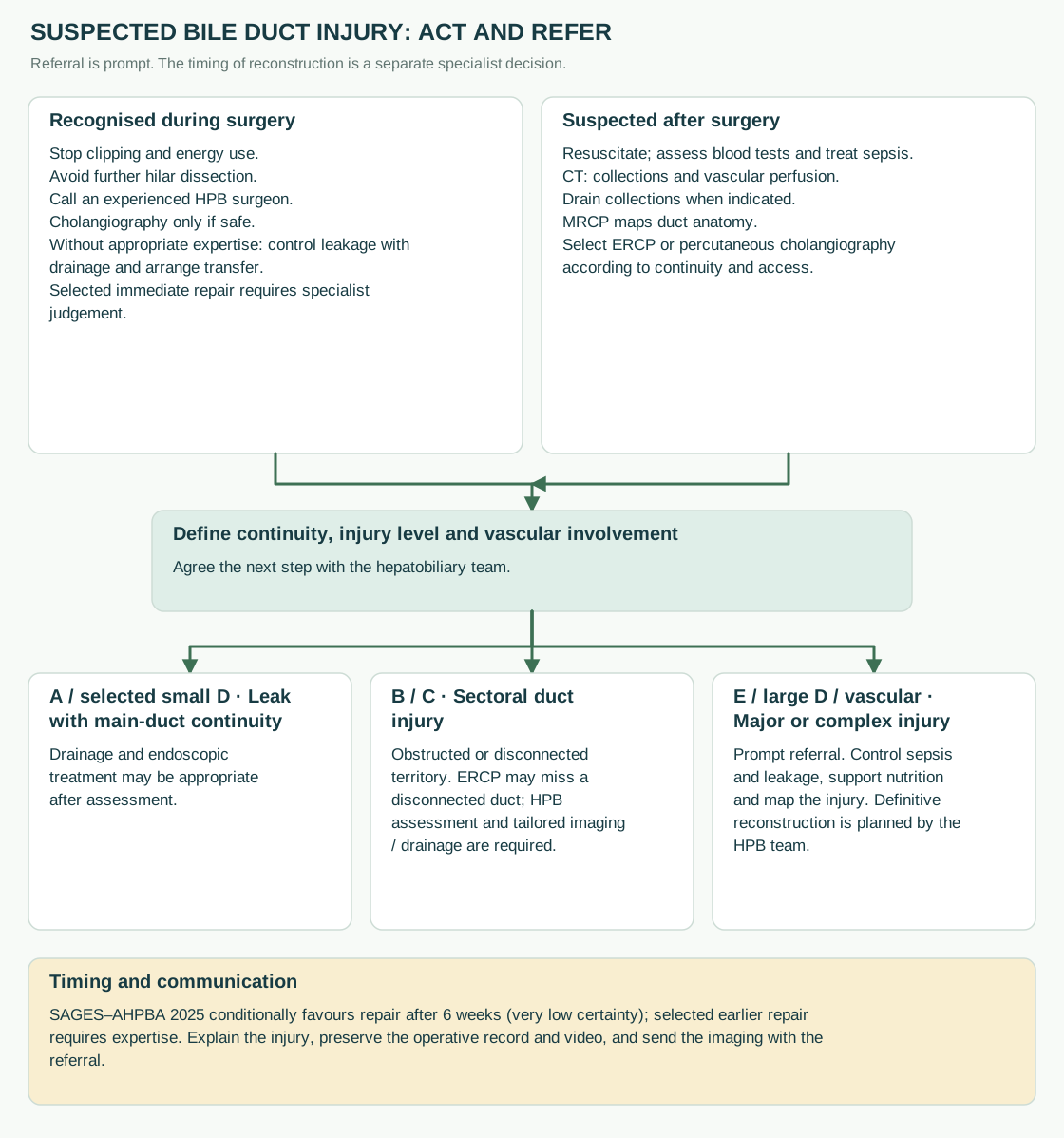
Diagram sources
Sources
Meta-analysis of 21 observational studies, 2,484 patients: reconstruction between 2 and 6 weeks should be avoided (Schreuder et al., BJS Open 2020); 2024 meta-analysis of 3,421 hepaticojejunostomies, same conclusion (Hiremath, Cureus 2024). WSES timing recommendations: WSES 2021. SAGES–AHPBA 2025 guideline for the surgical management of bile duct injury following cholecystectomy (Calabrese et al., Surg Endosc 2025): delayed repair over early repair, conditional, very low certainty; temporising drainage and transfer for a patient seen within 6 weeks when no hepatobiliary expert is on site; either minimally invasive or open repair, and either hepaticojejunostomy or hepaticoduodenostomy, by an experienced surgeon; its own meta-analysis noted that many of the early repairs with poor results were done by general surgeons. The guideline does not define a 2-to-6-week window; that comes from the meta-analyses above. Vasculobiliary injury: Strasberg and Helton, HPB 2011 (analytical review; the share of major injuries with an arterial injury varies widely between series). Guideline Systematic review Contested
Evidence and guidance
WSES 2021 (7.3–7.4) permits selected early specialist repair and, for major injuries presenting between 72 hours and 3 weeks, describes reconstruction after a minimum of 3 weeks once the acute/subacute situation has resolved. The SAGES–AHPBA 2025 guideline instead suggests repair beyond 6 weeks (conditional, very low certainty). These are separate recommendations; neither replaces specialist assessment of sepsis, injury anatomy and tissue condition. WSES BDI guideline, 2021.
08The referral pathway
- Every hospital doing cholecystectomy needs a named route to a hepatobiliary unit, used on the day of recognition.
- Send the operation note and video, cholangiogram and CT images, drain output, liver tests and the classification.
- Transfer once stable, drains in place, not after a local attempt at repair.
- Without a regional network, the equivalent is a telephone call to the nearest surgeon with a biliary reconstruction practice.
09Long-term outcomes
Quality of life stays lower for years. Follow up for at least 10 years after a surgical repair (SAGES–AHPBA 2025): liver tests at every visit and imaging for any rise, because anastomotic strictures appear beyond the 2-to-5-year window too. Cholangitis after a repair is a stricture until imaged. Guideline
- Not every injury declares itself in the first week. A thermal injury or a partly occluding clip presents months later with jaundice, itching or cholangitis from a stricture.
- An occluded sectoral duct (Strasberg B) can stay silent for years and then show as recurrent cholangitis, or as a shrunken atrophic segment on imaging done for something else.
- A neglected stricture ends in secondary biliary cirrhosis and portal hypertension in a minority; liver transplantation is the last resort for the rare end-stage case.
- Any cholangitis or unexplained jaundice after a cholecystectomy, however long ago, needs MRCP and the question: was the duct injured?
Source
Booij et al., Ann Surg 2018. Length of follow-up: SAGES–AHPBA 2025 (at least 10 years after surgical treatment).
10What examiners ask
- Draw the Strasberg classification and explain the difference between B and C.
- Bile in the field after dividing a structure: stop, help, cholangiogram, drain, do not repair, refer, tell the patient.
- Jaundice on day 3: liver tests, CT, then MRCP; ERCP only if a leak with continuity is likely.
- When to repair a major injury, and why not at 3 weeks.
- Why a right hepatic artery injury matters.
- A clip has gone on the common duct and you notice before anything is divided. What now?
- Define a bile leak, and grade it.
- The medicolegal consequences and the duty of candour (medicolegal issues; consent and communication).
Test yourself further: Practise questions on Bile duct injury in the Exam zone.