ICG fluorescence cholangiography
Indocyanine green shows you where the ducts are. It does not show you what is inside them, and it cannot replace dissection.
Reviewed against its sources on 3 November 2026. Open checks: the 2025 ICG meta-analysis figures come from its abstract.
- ICG shows where the extrahepatic ducts run; it does not show what is inside them or penetrate a thick inflamed triangle.
- Give it hours before, or a low dose at induction; in DOTIG timing mattered more than dose.
- Trials show earlier, better visualisation but cannot prove fewer injuries; ICG is an adjunct to the CVS, never a substitute.
01Mechanism
- Indocyanine green (ICG) binds plasma proteins, is taken up by hepatocytes and is excreted unchanged into bile.
- It absorbs at about 800 nm and fluoresces at about 830 nm: near-infrared, invisible to the eye, shown by a special camera as a green overlay. This is near-infrared fluorescence cholangiography.
- It shows where bile flows: the common duct, common hepatic duct, cystic duct and an unobstructed gallbladder. It does not show stones or the inside of the duct.
- The dye is cheap (a 25 mg vial costs a few pounds or dollars). The camera is the cost; most modern towers offer one.
02Dose, route and timing
Contested There is no agreed standard. These are the regimens used in the main trials. The one consistent finding: a longer gap between dose and imaging gives a better duct-to-liver picture, because the liver glows for the first hour or more.
| Regimen | Where used | Comment |
|---|---|---|
| 2.5 mg IV at least 30 to 45 min before incision | FALCON trial; many European centres | Simple. Bright liver in the first hour can hide the hilar ducts |
| 0.05 mg/kg IV during the operation | Danish non-inferiority trial (Lehrskov 2020) | Weight-based, low dose, given intra-operatively, so the liver is still bright; the trial compared it with X-ray cholangiography, not with earlier dosing |
| 2.5 mg or 0.05 mg/kg, more than 3 h or 15 to 30 min before (four arms) | DOTIG, 200 patients, Spain | More than 3 hours before beat dosing at induction. Timing mattered more than dose |
| 5 mg or 10 mg IV at least 3 h before imaging | Dutch optimisation study (Boogerd 2017) | Best duct-to-liver ratio 3 to 7 h after 5 mg and 5 to 25 h after 10 mg; liver as bright as the cystic duct for up to 3 h (5 mg) or 5 h (10 mg). Dosing the evening before needs the dose given on the ward or at pre-assessment |
| 0.3 mg/kg IV 6 h before induction | Greek three-arm trial (Symeonidis 2026) | Good visualisation, no duct injuries in 80 patients |
| Intracholecystic: a few mL of dilute ICG into the gallbladder after port placement | Same Greek trial; case series | No liver background; works within minutes; needs a puncture, may spill; dose reporting inconsistent |
- Safe in renal impairment (hepatic excretion). Allergic reactions are rare but real. Crosses the placenta minimally; limited pregnancy data.
- The US label (January 2026) has one contraindication: previous hypersensitivity to ICG. Maximum total dose 2 mg/kg, a ceiling not a target. The vial contains up to 5 percent sodium iodide; the label does not list iodine allergy as a contraindication.
- Contested The WSES 2025 emergency consensus says ICG is contraindicated with known iodine or contrast allergy. The two sources disagree.
- Practical position: a previous reaction to ICG is absolute. Iodine or contrast allergy means read the leaflet of the product in your hospital, discuss with the anaesthetist and have resuscitation drugs drawn up. The leaflet in the box, not the US label, applies to your patient.
More detail and sources
FALCON regimen: van den Bos et al., Surg Endosc 2023. Danish trial: Lehrskov et al., Br J Surg 2020 (0.05 mg/kg given intra-operatively). DOTIG: López-Sánchez et al., Surg Endosc 2025. Dose and dosing-time optimisation: Boogerd and colleagues reviewed the literature and measured bile duct-to-liver fluorescence ratios after 5 mg and 10 mg; administering 5 mg at least 3 hours before imaging was practical and gave ratios above 1.0 (Boogerd et al., Surg Innov 2017). Weight-based “evening before” regimens of 0.25 to 0.5 mg/kg are quoted in several reviews but were not re-verified against a primary series for this site, so no figure is given for them here. Greek trial: Symeonidis et al., Medicina 2026.
The EAES consensus on fluorescence-guided surgery strongly recommends ICG during laparoscopic cholecystectomy for better anatomical definition, without fixing a single dose (Cassinotti et al., Surg Endosc 2023). Guideline
The label and the guidance. The current United States prescribing information for IC-GREEN (revised January 2026) has one contraindication: a history of hypersensitivity to indocyanine green. It warns that hypersensitivity reactions including anaphylaxis, urticaria and deaths from anaphylaxis have been reported, and that resuscitation staff and equipment must be at hand. The maximum total dose is 2 mg/kg. The vial contains up to 5 percent sodium iodide, mentioned only because it can reduce thyroid uptake of radioactive iodine for about a week (IC-GREEN prescribing information, DailyMed, January 2026). Product label The 2025 WSES consensus on fluorescence-guided surgery in the emergency setting states that ICG is contraindicated in patients with known allergy to iodine or iodinated contrast agents (De Simone et al., World J Emerg Surg 2025). Contested Formulations and warnings differ between countries.
03How to interpret the image
- Before dissection: switch to fluorescence after placing the landmarks. In a thin patient you see the common duct and common hepatic duct in the ligament, often with the cystic duct joining. This is the map: where the main duct lies relative to the neck, and whether the cystic duct is short, long or joins unusually.
- During dissection: check repeatedly. Aim to see the cystic duct in continuity with the gallbladder and its junction with the common hepatic duct, with no unexplained fluorescent structure in the triangle.
- Before clipping: confirm that the structure to be clipped runs into the gallbladder, and that a fluorescent duct continues above and below it.
- Arteries: a second intravenous dose during the operation shows the arterial phase for 30 to 60 seconds and can confirm the cystic artery; the FALCON centres used this.
- Leak check: at the end, fluorescence in the bed or around the clips suggests a leak.
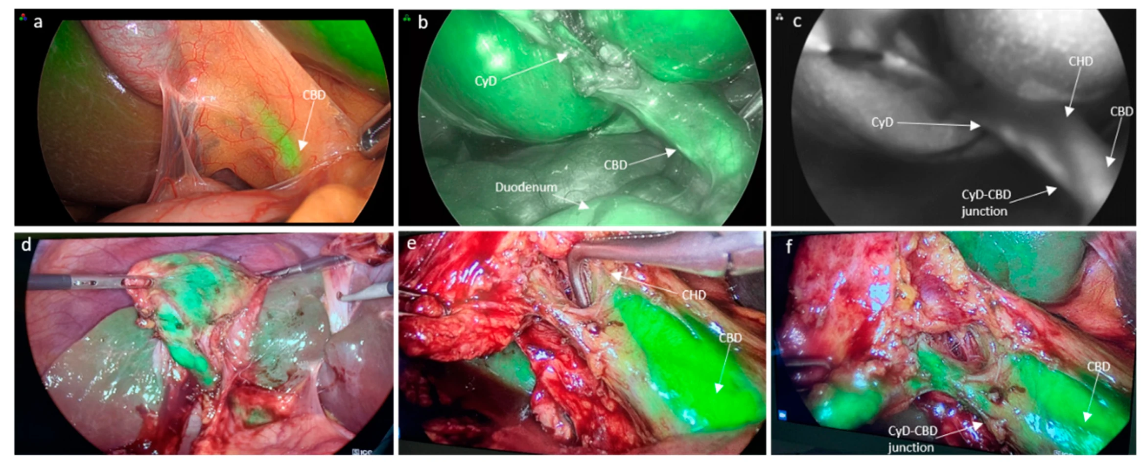
Image source and reuse
04What it cannot show
Stones
ICG does not image the lumen. A stone, filling defect or stricture needs X-ray cholangiography or ultrasound.
Deep structures
Near-infrared light penetrates only 5 to 10 mm. In acute cholecystitis, obesity and a fibrotic triangle, fat and oedema block the signal. The ducts are often unseen before dissection, exactly when you most want them.
An obstructed duct
A cystic duct blocked by a stone, or an aberrant duct draining an obstructed segment, does not fluoresce.
The liver background
Early after a dose the liver glows and hides the hilar ducts. Later dosing or intracholecystic injection avoids this.
Judgement
Fluorescence shows a green tube. It does not tell you what the tube is.
Every guideline: an adjunct to the CVS, never a substitute
Sources
Brunt et al., 2020; de’Angelis et al., 2021. Guideline The SAGES imaging guideline notes that fluorescence may be hard to see in acute cholecystitis.
05What the trials show
- Use ICG to help identify biliary anatomy; trials support better visualisation of some structures.
- Do not rely on ICG to avoid conversion: change strategy when safe progress is not possible.
- ICG does not establish CVS or make an unsafe dissection safe. A reduction in bile duct injury has not been established in randomised trials.
| Study | Design | Main finding |
|---|---|---|
| Dip 2019 | RCT, 639 patients, ICG plus white light versus white light | Structures identified more often before and after dissection; no difference in injury (underpowered) |
| Lehrskov 2020 | RCT non-inferiority, 120, ICG versus X-ray | Junction seen 49/60 versus 51/60; non-inferior; 2 versus 4.8 minutes |
| FALCON 2023 | RCT, 294 elective, six European centres | CVS 4 minutes faster; cystic duct at 6 versus 13 minutes; no difference in total time, complications or conversion |
| DOTIG 2025 | RCT, 200, four dose-and-timing arms | Dye more than 3 hours before gave better images than at induction; better images went with shorter operations and fewer complications (association) |
| Pimentel 2025 | Meta-analysis of 8 RCTs, 1,586 patients | Better identification; injury OR 0.73 (95% CI 0.05 to 10.80): compatible with benefit, no effect or harm |
| Symeonidis 2026 | RCT, 240, X-ray versus IV versus intracholecystic ICG | Both ICG routes non-inferior, faster (about 2 versus 5 minutes), easier; three X-ray failures; no injuries |
| Neel 2026 | Cohort, 1,266,024 US operations 2016 to 2024, weighted | Injury 0.25 versus 0.40 percent; conversion 0.40 versus 0.84; fewer biliary interventions. Association, not proof |
| Haverinen 2025 | Retrospective, 2,009, Swedish fundus-first unit with routine X-ray | Conversion 1.2 versus 3.3 percent; no difference in injury (0.4 versus 0.5) |
Sources
Dip et al., Ann Surg 2019; Lehrskov et al., Br J Surg 2020; van den Bos et al., Surg Endosc 2023 (FALCON); López-Sánchez et al., Surg Endosc 2025 (DOTIG); Pimentel et al., Surgery 2025 (figures as reported in the abstract); Symeonidis et al., Medicina 2026; Neel et al., JAMA Surg 2026 (relative risk 0.62, 95 percent confidence interval 0.49 to 0.79; surgeons who adopt ICG may differ from those who do not); Haverinen et al., Front Surg 2025. Contested
06ICG compared with X-ray cholangiography
| ICG fluorescence | X-ray cholangiography (IOC) | |
|---|---|---|
| Shows | Course of the ducts, from outside | The lumen: anatomy, stones, strictures, leaks, flow to duodenum |
| When | Before any dissection and repeatedly | Only after the cystic duct is identified and opened |
| Time | Seconds; about 2 minutes in trials | 5 to 15 minutes; radiographer, fluoroscopy, catheter |
| Fails | In inflammation and obesity | Cannulation fails in 5 to 10 percent |
| Radiation, contrast | None | Both; shielding in pregnancy |
| Duct stones | No | Yes, with transcystic clearance in the same sitting |
| Injury once it has happened | May show a leak or interrupted duct | Shows the level precisely; test of choice |
| Guideline position | Adjunct to the CVS (all); strongly recommended by EAES 2023; not routine per WSES 2021 | Liberal use in acute disease and unclear anatomy (2020 consensus); routine suggested over selective, and IOC over ICG, by SAGES 2025 (conditional) |
ICG fluorescence
- Shows
- Course of the ducts, from outside
- When
- Before any dissection and repeatedly
- Time
- Seconds; about 2 minutes in trials
- Fails
- In inflammation and obesity
- Radiation, contrast
- None
- Duct stones
- No
- Injury once it has happened
- May show a leak or interrupted duct
- Guideline position
- Adjunct to the CVS (all); strongly recommended by EAES 2023; not routine per WSES 2021
X-ray cholangiography (IOC)
- Shows
- The lumen: anatomy, stones, strictures, leaks, flow to duodenum
- When
- Only after the cystic duct is identified and opened
- Time
- 5 to 15 minutes; radiographer, fluoroscopy, catheter
- Fails
- Cannulation fails in 5 to 10 percent
- Radiation, contrast
- Both; shielding in pregnancy
- Duct stones
- Yes, with transcystic clearance in the same sitting
- Injury once it has happened
- Shows the level precisely; test of choice
- Guideline position
- Liberal use in acute disease and unclear anatomy (2020 consensus); routine suggested over selective, and IOC over ICG, by SAGES 2025 (conditional)
Complementary, not rivals. ICG is a map that works before the first cut; cholangiography is a test that works once you have found the cystic duct. Laparoscopic ultrasound is a third option in experienced hands; SAGES considers it equivalent to cholangiography. A laparoscopic probe run along the hepatoduodenal ligament shows the common duct in cross-section, the portal vein and hepatic artery behind it, and a stone as a bright echo with a shadow. It needs no cannulation, contrast or radiation, so it can be done before any dissection and repeated, but it is operator dependent and the probe and the skill are uncommon outside units that use it routinely. Expert opinion
Changed recently The 2025 suggestion of routine cholangiography is conditional, on low or very low certainty observational evidence, and calls for shared decision-making. With normal liver tests, a definitive CVS and no other indication, routine cholangiography “could be considered on an individual basis, taking into account surgeon experience, patient-specific factors, and institutional resources”. Fluoroscopy licensing and C-arm availability are legitimate constraints; clinical autonomy is “preserved and expected”. The preference for cholangiography over ICG is graded the same way. The guideline does not make an operation without cholangiography substandard, and does not tell a unit that uses ICG well to stop.
Sources
07How to perform and read an intra-operative cholangiogram
Doing it
- Set up before you need it. Catheter, contrast diluted about half and half with saline, a three-way tap with a saline and a contrast syringe, the C-arm in the room and the radiographer warned at the time-out; nothing radio-opaque under the right upper quadrant. Expert opinion
- In the routine case, after the Critical View. Clip the cystic duct at the gallbladder end, open its front wall with scissors a short distance from the gallbladder, and milk the duct towards the gallbladder to empty bile and small stones. When the anatomy will not resolve, image through the gallbladder instead (bail-out) and never open or cannulate a structure you cannot name.
- Cannulate. Pass the catheter through a lateral port or a separate stab, into the cystic duct for a short distance and never to the junction; hold it with a cholangiogram clamp or a loose clip; flush with saline and clear every bubble from the line before contrast goes in.
- Image. A small test dose, then a steady fill under fluoroscopy; tilt the table or roll the patient so the duct clears the spine; instruments out of the field; save the images.
- No flow into the duodenum? Give glucagon or hyoscine and repeat. An impacted stone or a clip on the common duct does not open with either. If the right system does not fill, do not go on until it is explained.
- Finish. Withdraw the catheter, clip the cystic duct below the opening on healthy duct, and leave a transcystic wire in place if a rendezvous ERCP is planned. Cannulation fails in about 5 to 10 percent; a stone found leads to the clearance pathway.
Reading it
Read it systematically, before anything else is divided. It takes a minute. Expert opinion
- Technique first. Catheter in the cystic duct, not through it; no spill; table tilted so the duct is not behind the spine; no air (a bubble is round, moves and rises; a stone is fixed and often faceted).
- Proximal filling: both hepatic systems. Right and left ducts and the right anterior and posterior sectoral ducts all fill. A missing right posterior duct is the finding that matters most: a low sectoral duct that has not filled, a clip on it, or an injury. Do not go on until it is explained.
- Cystic duct and junction. Length, how it joins (lateral, spiral, low, parallel), and that the cannulated structure is the cystic duct, not the common duct.
- Common duct. Calibre (normal under about 6 to 8 mm), filling defects (stone, air, tumour), tapered or abrupt cut-off (stricture, clip, impacted stone), extravasation.
- Distal flow into the duodenum. No flow means an impacted stone, sphincter spasm (glucagon or hyoscine, repeat) or a clip on the common duct.
- Say it and write it. “Both hepatic systems fill, cystic duct joins laterally, common duct 5 mm, no filling defect, free flow to the duodenum.” Any item not ticked: stop, nothing clipped or divided, senior help.
Two patterns on sight. A filling defect below the junction with a dilated duct and slow flow is a stone: transcystic clearance or ERCP (clearance pathway). A cut-off with no proximal filling, or contrast in the field, is an injury until proved otherwise (bile duct injury).
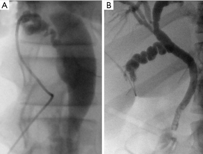
Image source and reuse
Source
Technique and reading sequence after the SAGES clinical spotlight reviews on cholangiography and duct exploration, and the practice of units using cholangiography routinely (SAGES clinical spotlight review).
08Cost and availability
- The dye costs little. The camera system costs tens of thousands, so uptake is uneven: standard in many European and North American centres and large Indian corporate hospitals; uncommon in district hospitals and most public-sector units in low- and middle-income countries.
- Without ICG nothing changes in the method of identification: the CVS, liberal X-ray cholangiography and a low threshold for bail-out remain the standard. An operation without ICG is not an unsafe operation.
09What examiners ask
- Mechanism, dose and timing, and why dosing the night before gives a better picture.
- Two things ICG cannot do: show stones; penetrate an inflamed thick neck.
- The evidence in one sentence: better and earlier visualisation in trials; an association with fewer injuries in a 1.27-million-patient cohort in 2026; no randomised proof of fewer injuries.
- ICG and the CVS: adjunct, not substitute.
- What SAGES 2025 suggests, and what the June 2026 clarification says about applying it.
- Read this cholangiogram: the six steps, and what a missing right posterior duct means.
Test yourself further: Practise questions on ICG fluorescence in the Exam zone.